Copyright
©The Author(s) 2021.
World J Clin Cases. Dec 26, 2021; 9(36): 11448-11456
Published online Dec 26, 2021. doi: 10.12998/wjcc.v9.i36.11448
Published online Dec 26, 2021. doi: 10.12998/wjcc.v9.i36.11448
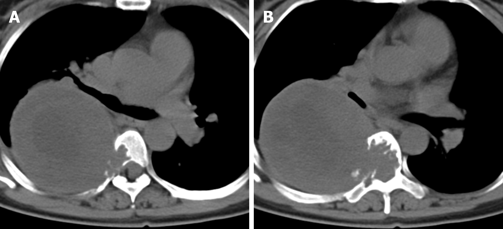
Figure 1 Axial computed tomography revealed bone destruction visible in the T5 (A) and T6 (B) vertebrae and the right accessory and part of the right rib, and irregular soft tissue density shadows were seen in the T5 and T6 vertebrae, which protruded into the thoracic cavity to the right.
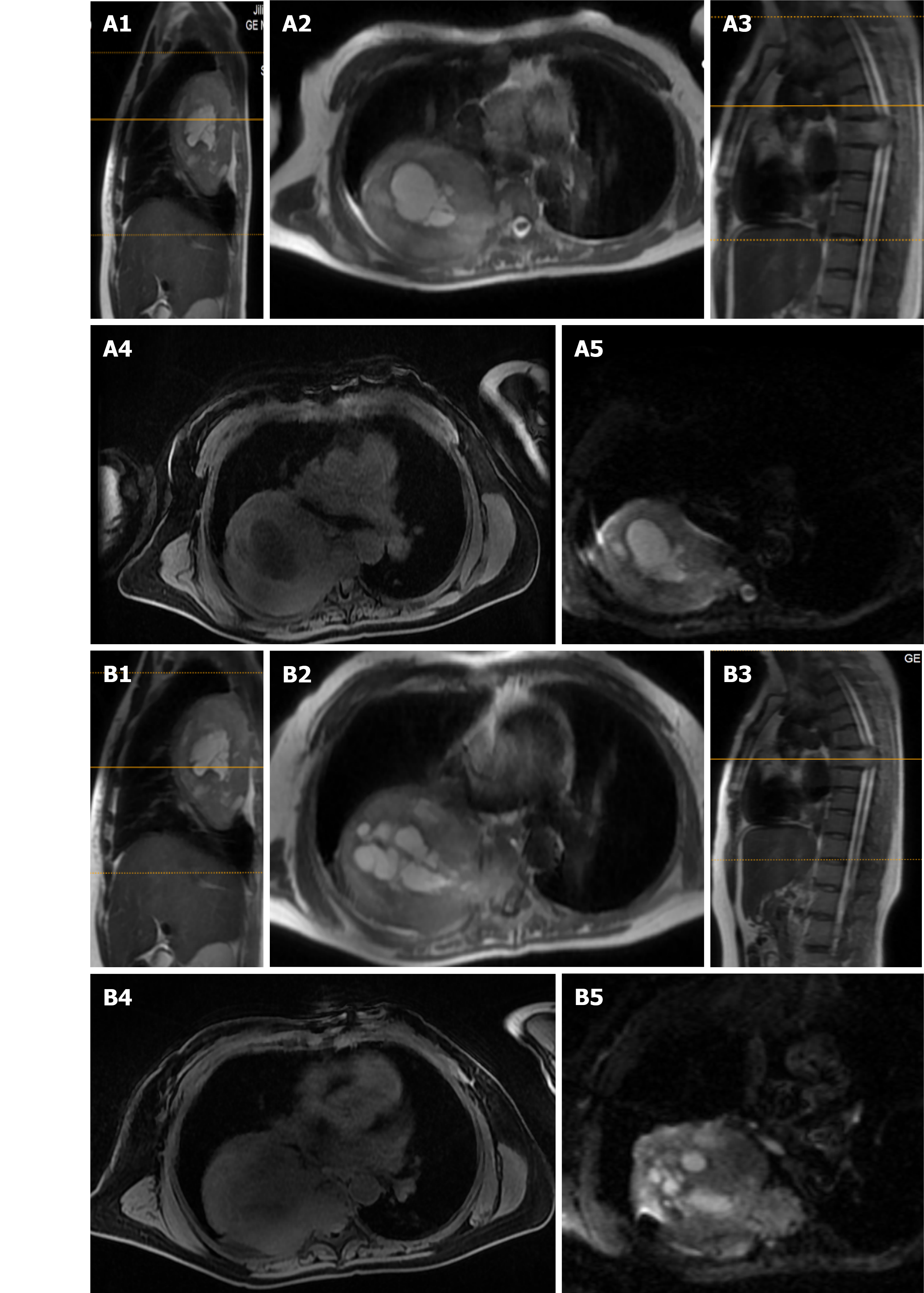
Figure 2 Sagittal and axial magnetic resonance imaging revealed tumor invasion at T5 (A1-4) and T6 (B1-4), and diffusion weighted imaging showed patchy high signals within the T5 (A5) and T6 (B5) lesions.
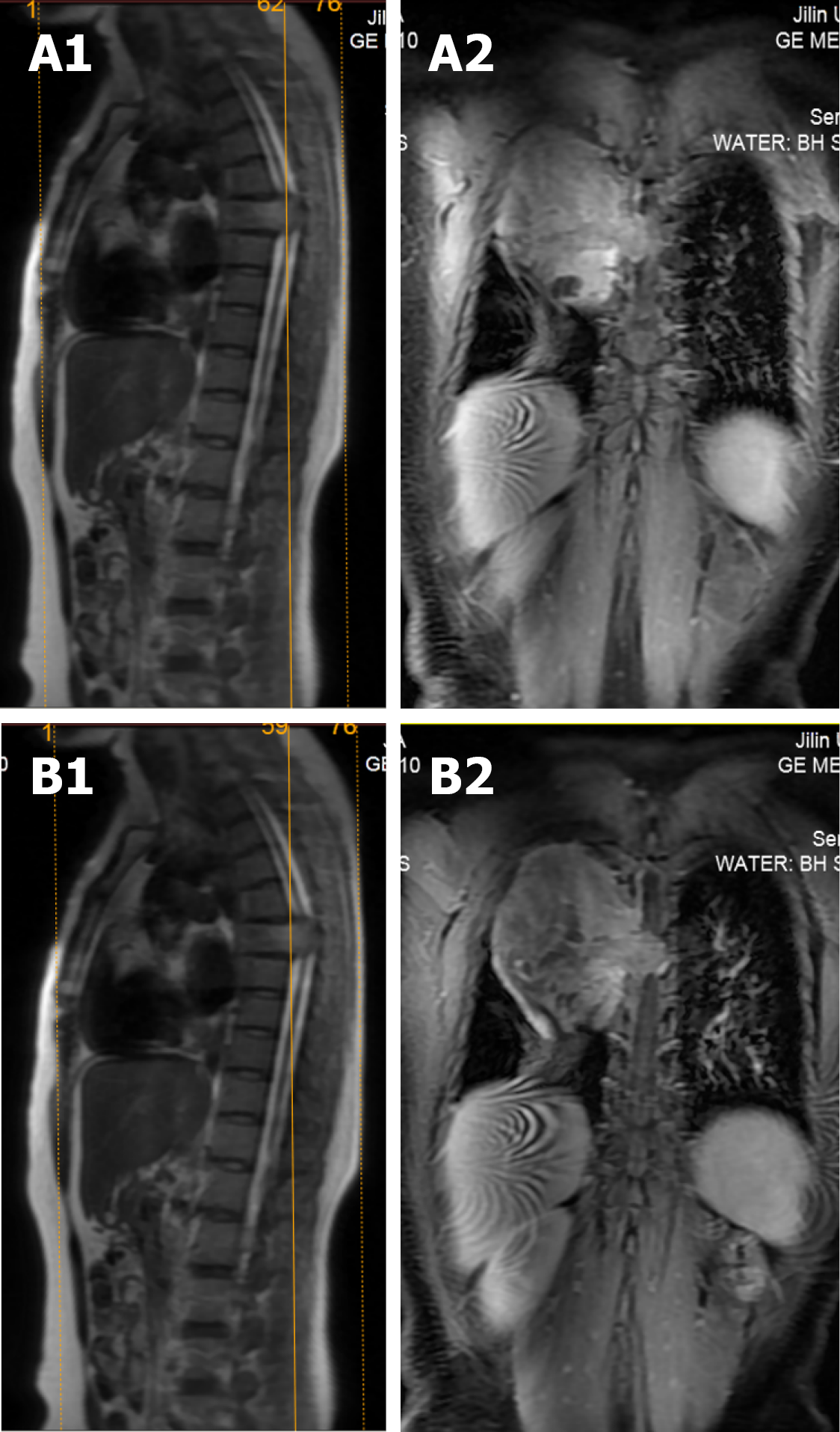
Figure 3 Sagittal and coronal magnetic resonance imaging revealed intravertebral occupancy at T6 (A1-2) and T5 (B1-2).
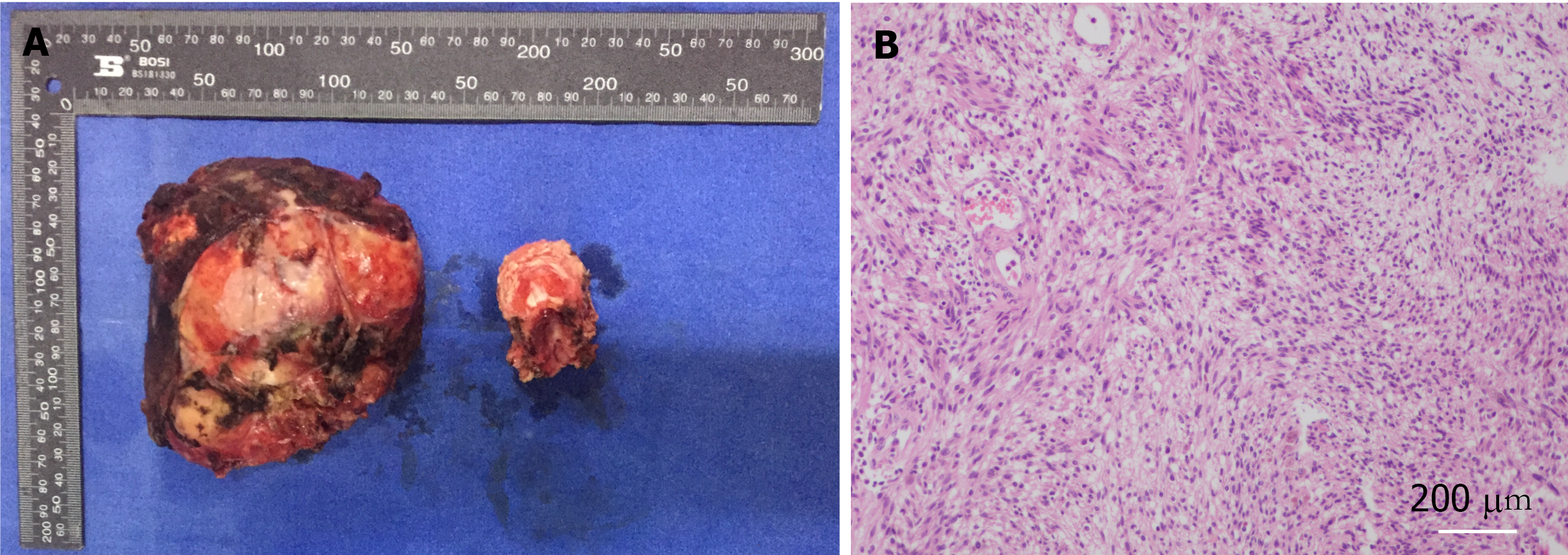
Figure 4 Histopathology of schwannomas.
A Perioperative resection of the tumor (left side) and vertebral bodies (right side); B shows a hematoxylin and eosin-stained section (×100 magnification) of the tumor.
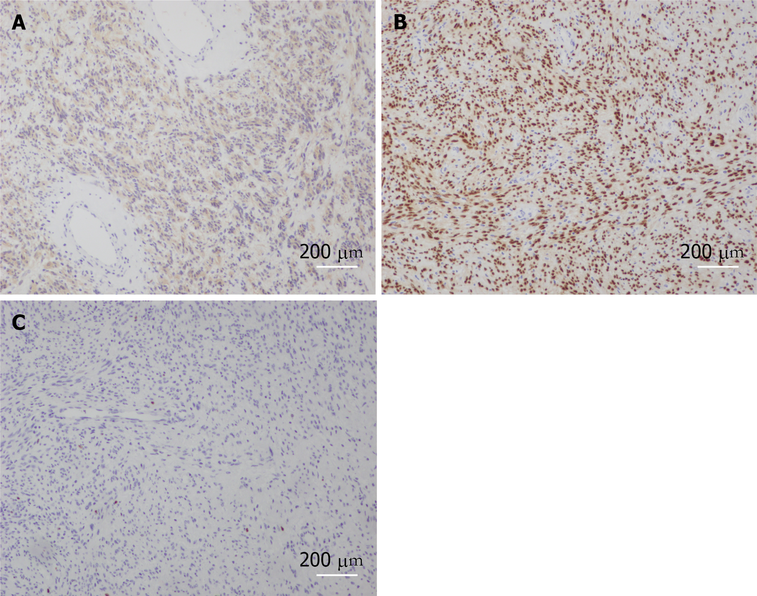
Figure 5 Histopathology of schwannomas.
A-C: Immunofluorescence staining of ‘the specific tissue’ for S-100, SOX10 and Ki67.
Figure 6 Nine months postoperative review, see internal fixation in place, no tumor recurrence found (A-C).
- Citation: Zhou Y, Liu CZ, Zhang SY, Wang HY, Nath Varma S, Cao LQ, Hou TT, Li X, Yao BJ. Giant schwannoma of thoracic vertebra: A case report. World J Clin Cases 2021; 9(36): 11448-11456
- URL: https://www.wjgnet.com/2307-8960/full/v9/i36/11448.htm
- DOI: https://dx.doi.org/10.12998/wjcc.v9.i36.11448


