Copyright
©The Author(s) 2021.
World J Clin Cases. Dec 26, 2021; 9(36): 11437-11442
Published online Dec 26, 2021. doi: 10.12998/wjcc.v9.i36.11437
Published online Dec 26, 2021. doi: 10.12998/wjcc.v9.i36.11437
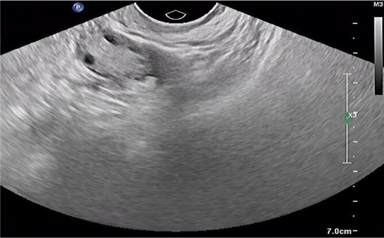
Figure 1 Vaginal ultrasound showed a mixed echogenic mass in the right ovary.
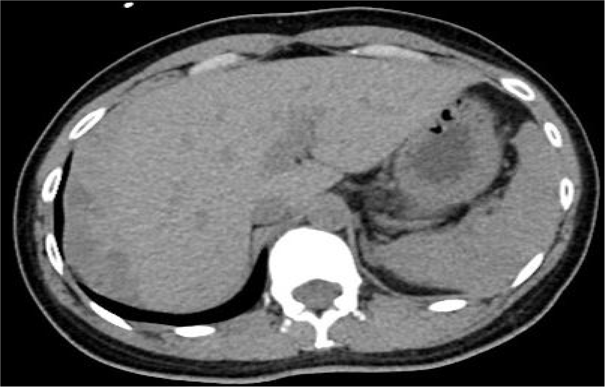
Figure 2 Computed tomography plain scan showed a curved high density mass beneath the subhepatic space.
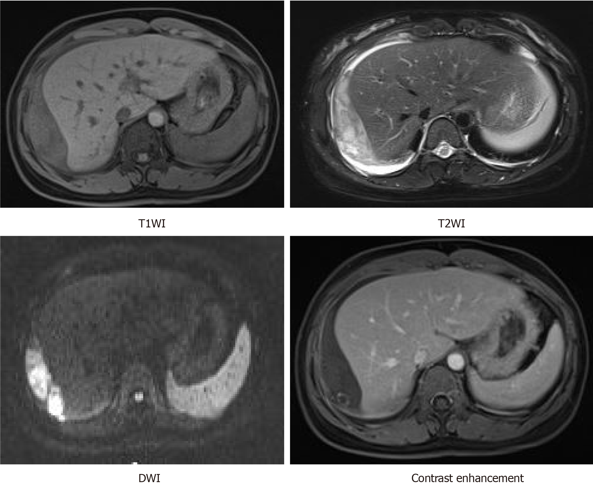
Figure 3 Magnetic resonance imaging scan showed a curved mixed signal beneath the subhepatic space, mostly low in T1-weighted imaging and high in T2-weighted imaging.
Diffusion-weighted imaging showed restricted diffusion. A peripheral enhanced nodule was observed within the mixed curved signal after administration of gadolinium. T1WI: T1-weighted imaging; T2WI: T2-weighted imaging; DWI: Diffusion-weighted imaging.
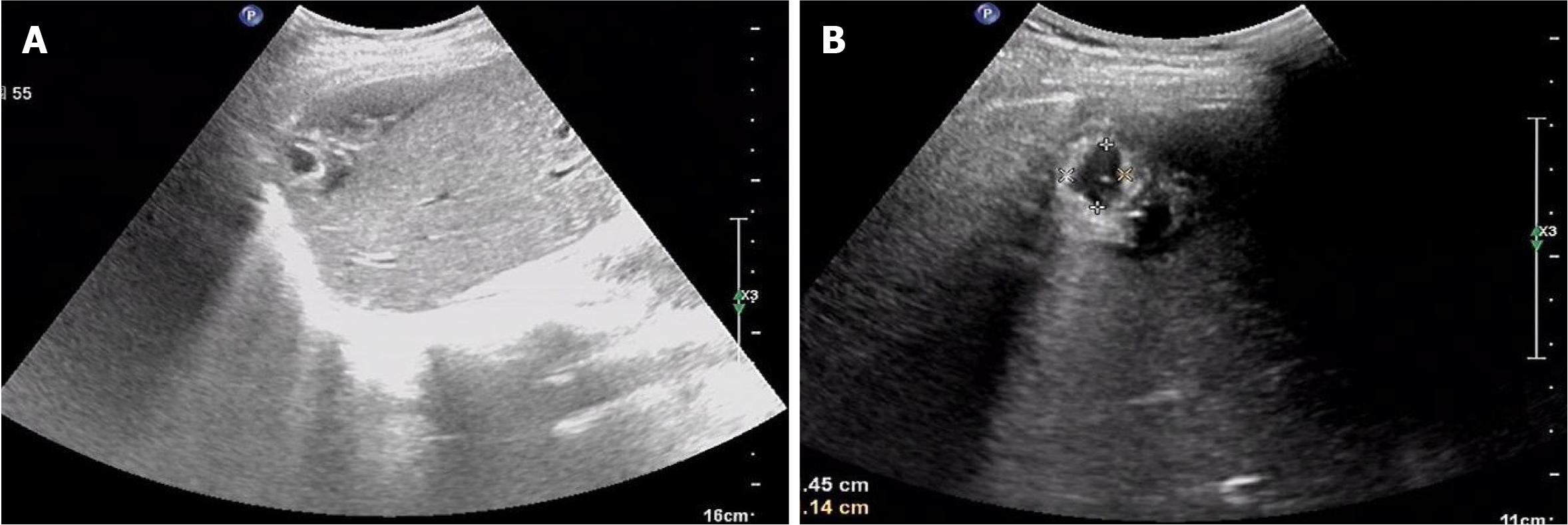
Figure 4 Abdominal ultrasound showed a mixed echogenic mass in the right lobe of the liver near the apex of the diaphragm.
A: The mass was approximately 1.5 cm × 1.1 cm in size; B: The mass showed characteristic features of a gestational sac, with a bud of approximately 0.4 cm in length (indicated by a bright dot in the dark zone).
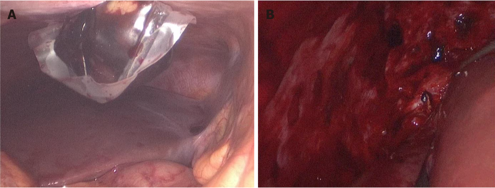
Figure 5 A mass measuring 5 cm × 3 cm, with dark red surface, was found under the right diaphragm on operation.
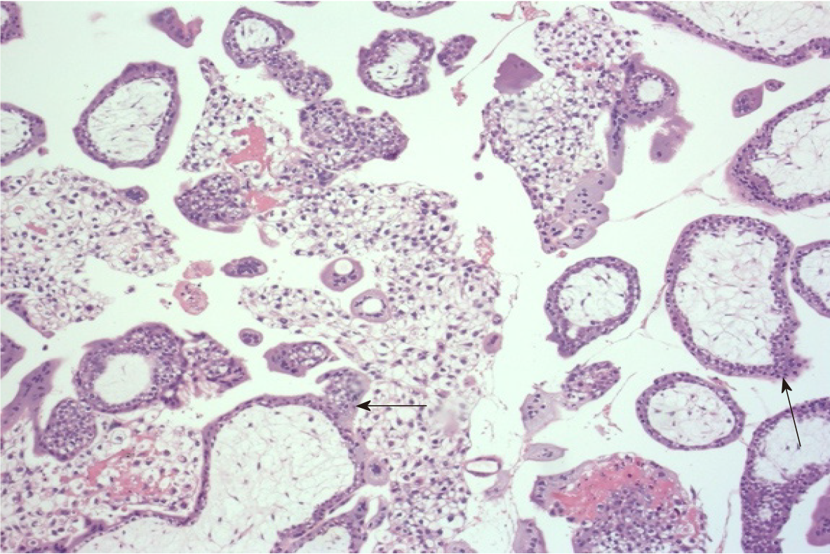
Figure 6 Histopathology findings confirmed the diagnosis of ectopic pregnancy.
Chorionic villi were present within the mass, with no features of abnormal trophoblastic proliferation (black arrows).
- Citation: Wu QL, Wang XM, Tang D. Ectopic pregnancy implanted under the diaphragm: A rare case report. World J Clin Cases 2021; 9(36): 11437-11442
- URL: https://www.wjgnet.com/2307-8960/full/v9/i36/11437.htm
- DOI: https://dx.doi.org/10.12998/wjcc.v9.i36.11437


