Copyright
©The Author(s) 2021.
World J Clin Cases. Dec 26, 2021; 9(36): 11419-11424
Published online Dec 26, 2021. doi: 10.12998/wjcc.v9.i36.11419
Published online Dec 26, 2021. doi: 10.12998/wjcc.v9.i36.11419
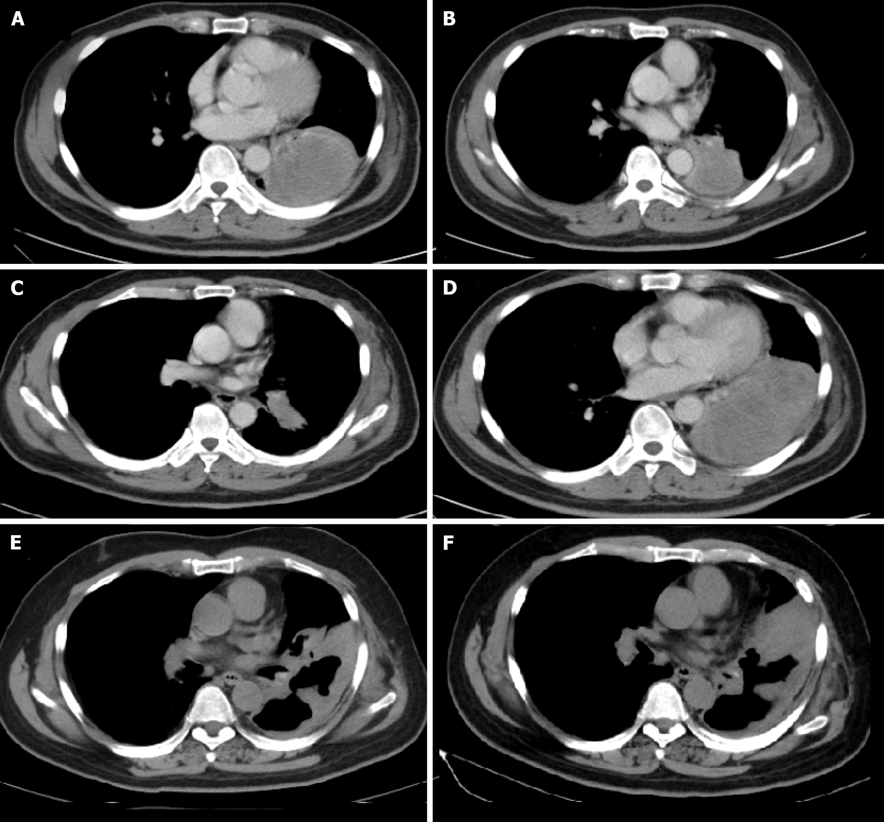
Figure 1 Computed tomography imaging of the non-small cell lung cancer patient.
A: Computed tomography imaging showed the mass located in lower lobe of left lung before gefitinib treatment; B: The mass enlarged and increased slightly after 11 mo of gefitinib treatment; C; The mass shrank significantly after treated with 2 cycles of pemetrexed plus carboplatin; D: The mass enlarged sharply after treated with 5 cycles of pemetrexed; E: The mass shrank significantly during pembrolizumab treatment; F: The mass enlarged and increased slightly after 10 mo of pembrolizumab treatment.
Figure 2 Timeline of events since the diagnosis and summary of administered treatments.
- Citation: Li D, Cheng C, Song WP, Ni PZ, Zhang WZ, Wu X. Dramatic response to immunotherapy in an epidermal growth factor receptor-mutant non-small cell lung cancer: A case report. World J Clin Cases 2021; 9(36): 11419-11424
- URL: https://www.wjgnet.com/2307-8960/full/v9/i36/11419.htm
- DOI: https://dx.doi.org/10.12998/wjcc.v9.i36.11419


