Copyright
©The Author(s) 2021.
World J Clin Cases. Dec 26, 2021; 9(36): 11392-11399
Published online Dec 26, 2021. doi: 10.12998/wjcc.v9.i36.11392
Published online Dec 26, 2021. doi: 10.12998/wjcc.v9.i36.11392
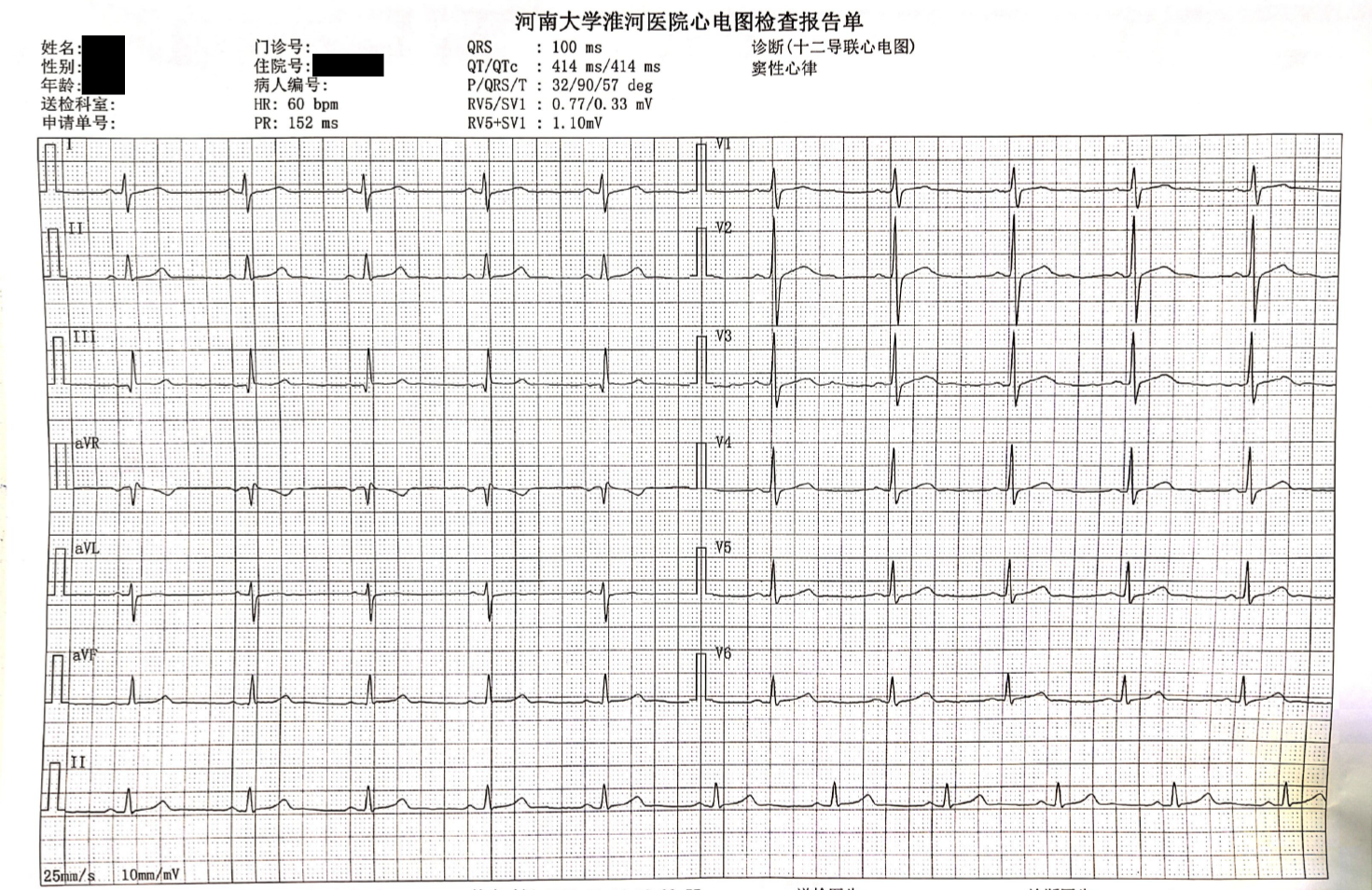
Figure 1 Electrocardiography.
Electrocardiogram tracings shows sinus rhythm with no arrhythmias.
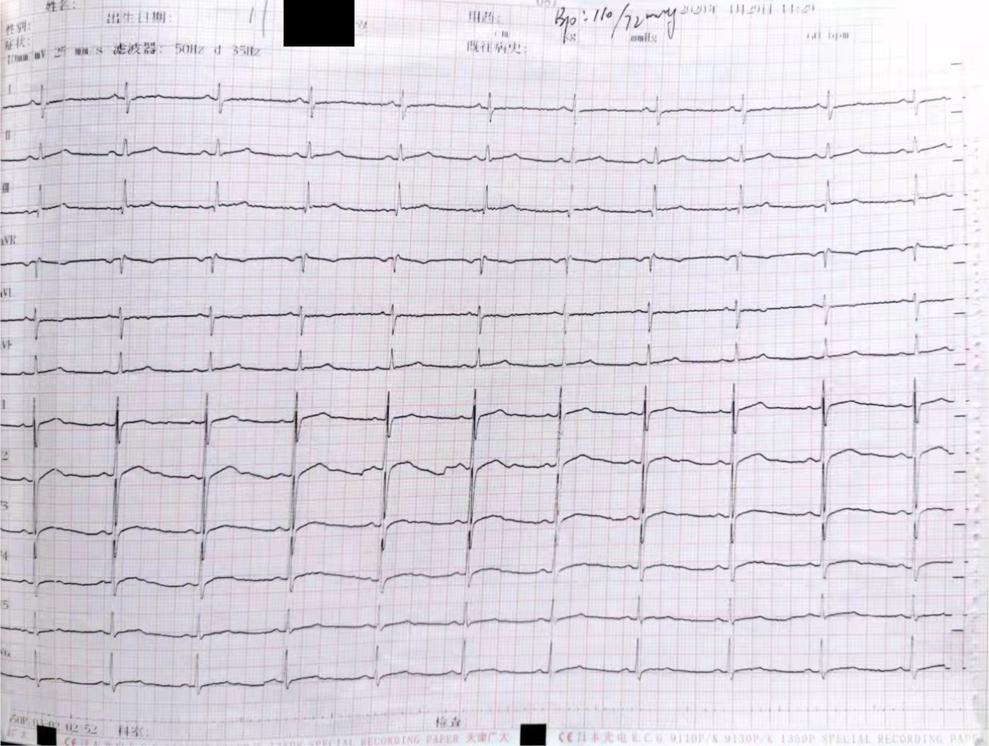
Figure 2 Electrocardiography.
Electrocardiogram tracings shows T wave is low on V4-6 limb lead.
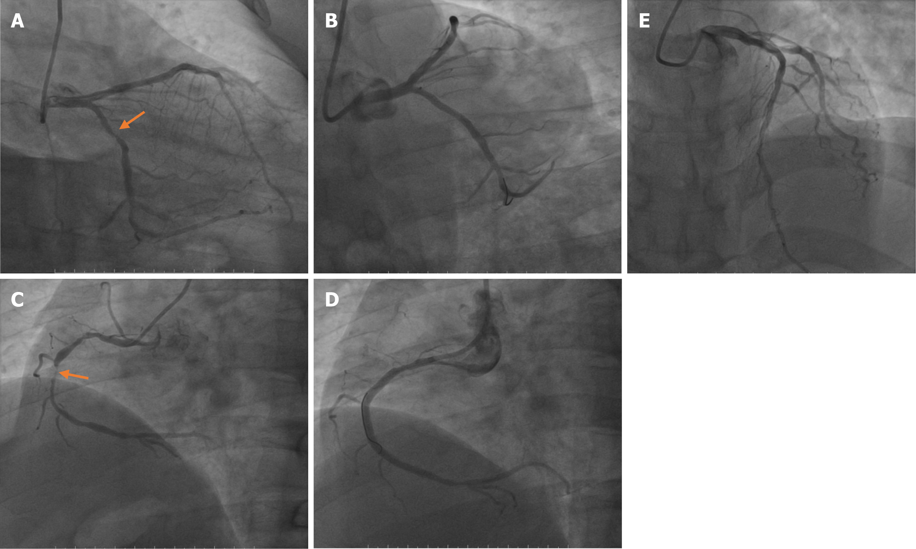
Figure 3 Surgical records of percutaneous coronary intervention.
A: Diffuse lesions in the near middle of left circumflex coronary artery (LCX). The most severe degree of stenosis is 80%; B: One 2.75 mm × 2 3 mm stent was placed in LCX; C: Diffuse lesions occurred throughout right coronary artery (RCA). The most severe degree of stenosis is 95%; D: Two 3.0 mm × 30 mm stents were placed in RCA; E: Multiple diseased plaques throughout left anterior descending coronary artery.
- Citation: Wan ZH, Wang J, Zhao Q. Acute myocardial infarction in a young man with ankylosing spondylitis: A case report. World J Clin Cases 2021; 9(36): 11392-11399
- URL: https://www.wjgnet.com/2307-8960/full/v9/i36/11392.htm
- DOI: https://dx.doi.org/10.12998/wjcc.v9.i36.11392


