Copyright
©The Author(s) 2021.
World J Clin Cases. Dec 26, 2021; 9(36): 11369-11381
Published online Dec 26, 2021. doi: 10.12998/wjcc.v9.i36.11369
Published online Dec 26, 2021. doi: 10.12998/wjcc.v9.i36.11369
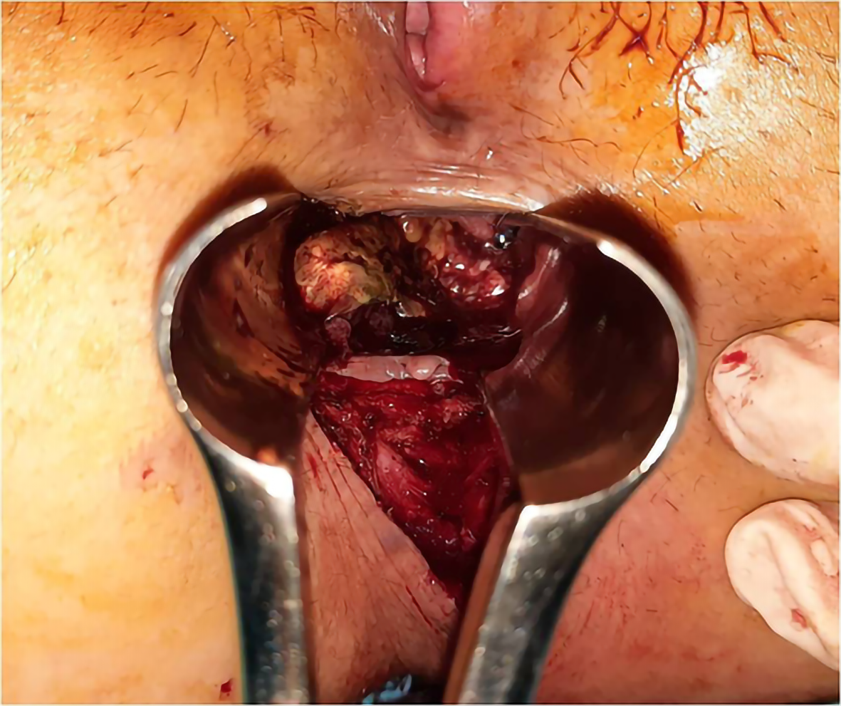
Figure 1 Aspect of the tumor at anoscopy.
Image shows a sessile tumor, near the anal verge, on the postero-lateral anorectal wall.
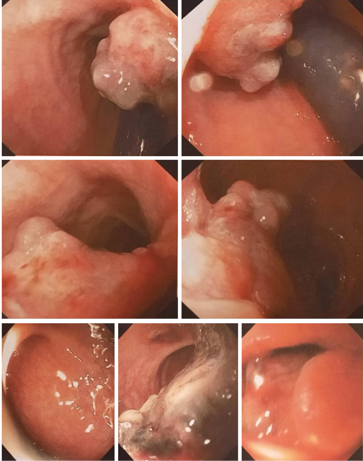
Figure 2 Colonoscopic aspect of the tumor.
Images show a cauliflower tumor with a wide base, exulcerated areas, and adherent clot, without obvious color changes or obstruction.
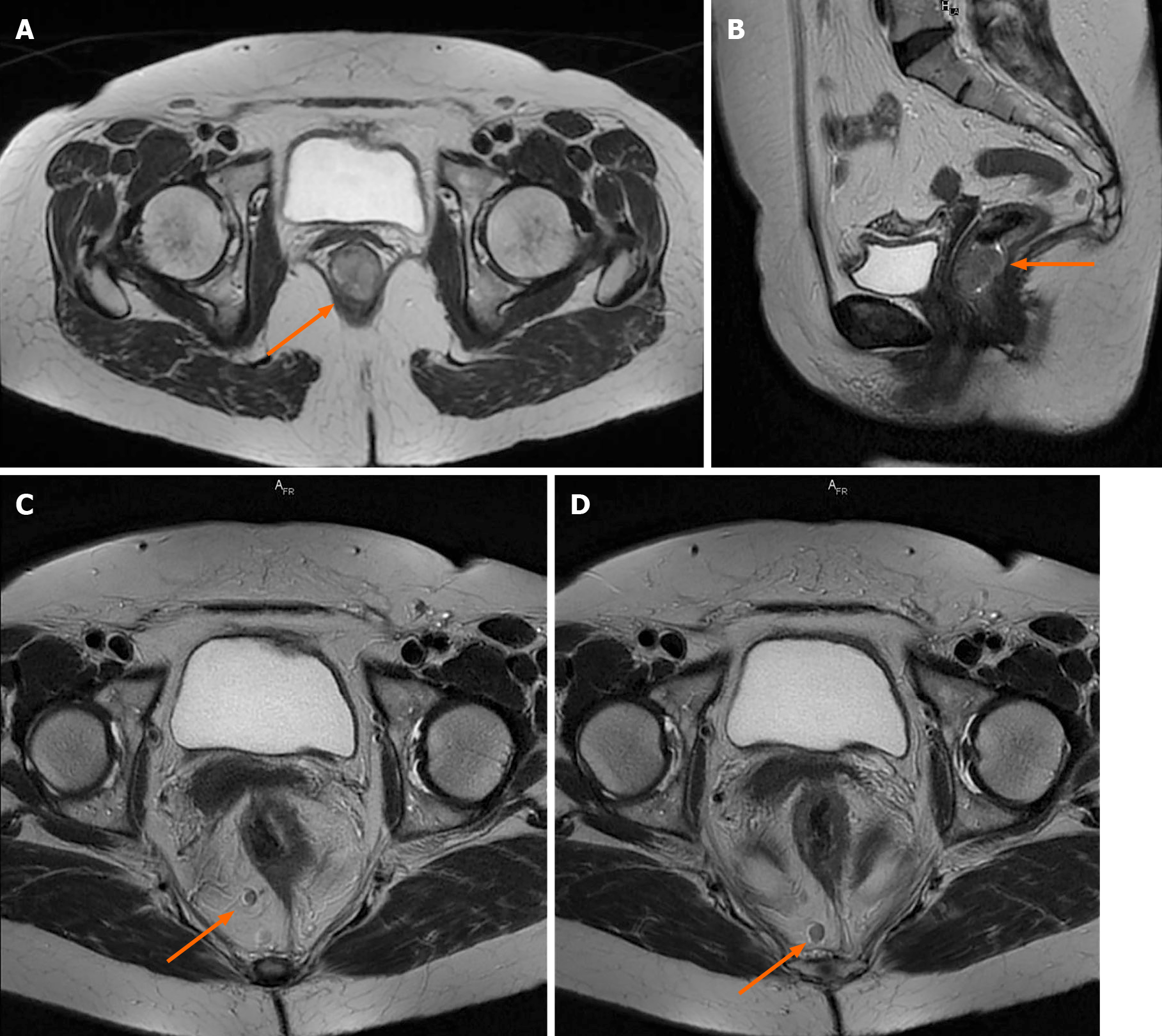
Figure 3 Pelvic magnetic resonance imaging scan with intravenous gadolinium administration.
A and B: Axial and sagittal T2-weighted images showing the tumor occupying the anorectal junction canal, with homogenous enhancement, without infiltration of the rectal fascia; C and D: Axial T2-weighted propeller images showing two large lymph nodes located in primary stations, without mesorectal fascia retraction.
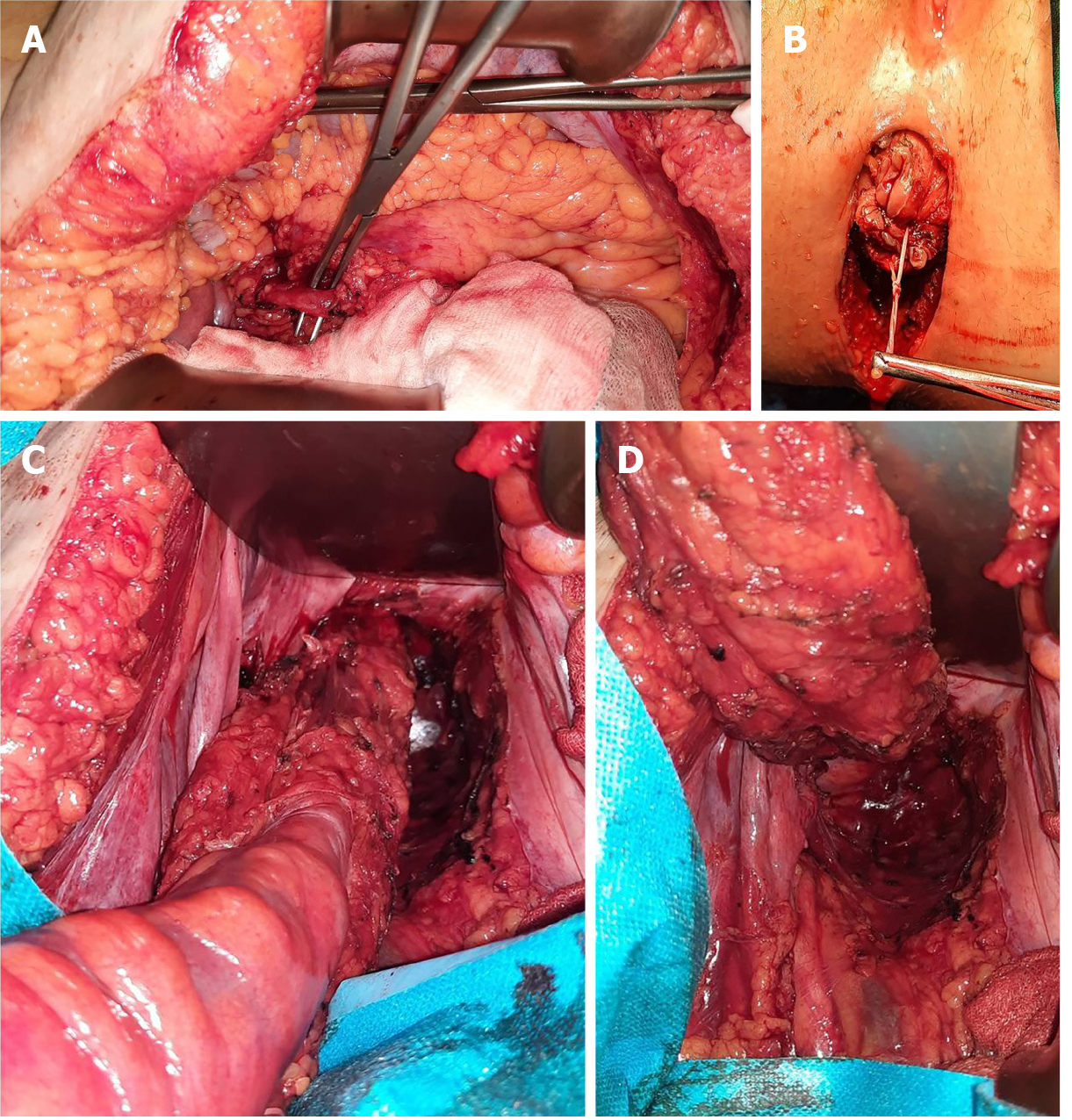
Figure 4 Abdominoperineal resection intra-operative images.
A: Dissection and identification of the inferior mesenteric artery; B: Perineal incision; C, D: Abdominal view of the dissected specimen, with total mesocolic and mesorectal excision.
Figure 5 Images of the resected specimen.
The tumor was identified adjacent to the dentate line with rectal extension.
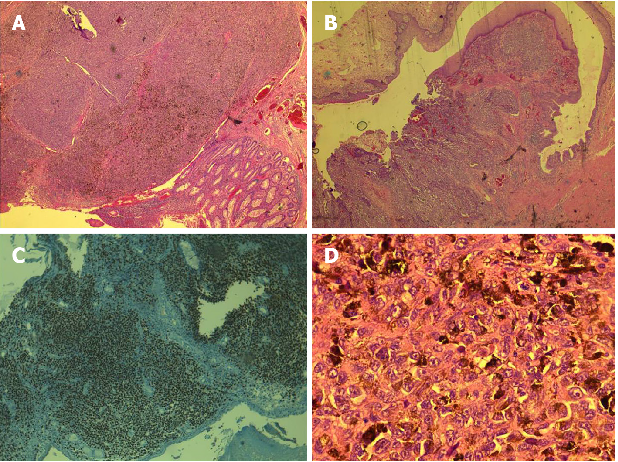
Figure 6 Histopathological and immunohistochemical examination of the tumor.
A: Immunohistochemistry image with SOX10 positivity; B and C: Tumoral infiltration confined to the submucosa, with pigmented areas, epithelioid cells, and localized fusiform aspect; D: Cells with eosinophilic cytoplasm, with melanocyte pigment.
- Citation: Apostu RC, Stefanescu E, Scurtu RR, Kacso G, Drasovean R. Difficulties in diagnosing anorectal melanoma: A case report and review of the literature. World J Clin Cases 2021; 9(36): 11369-11381
- URL: https://www.wjgnet.com/2307-8960/full/v9/i36/11369.htm
- DOI: https://dx.doi.org/10.12998/wjcc.v9.i36.11369


