Copyright
©The Author(s) 2021.
World J Clin Cases. Dec 26, 2021; 9(36): 11355-11361
Published online Dec 26, 2021. doi: 10.12998/wjcc.v9.i36.11355
Published online Dec 26, 2021. doi: 10.12998/wjcc.v9.i36.11355
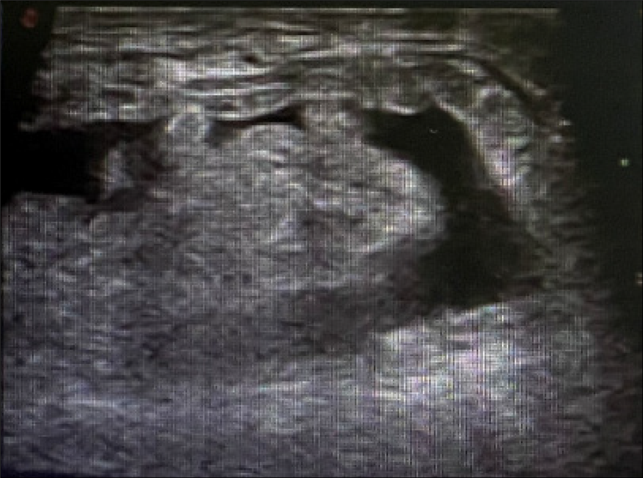
Figure 1 Inguinal B-ultrasound.
The right inguinal canal was involved, and there was no obvious reduction after pressurization with probes.
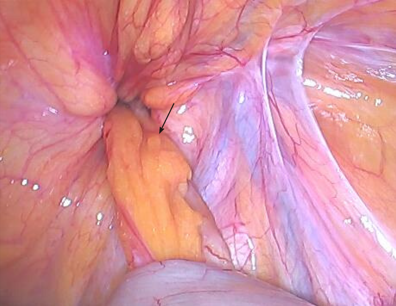
Figure 2 Appendix and femoral ring.
The arrow shows the appendix. The appendix and its mesangium were herniated into the femoral ring without reduction, and the color of the proximal segment was normal.
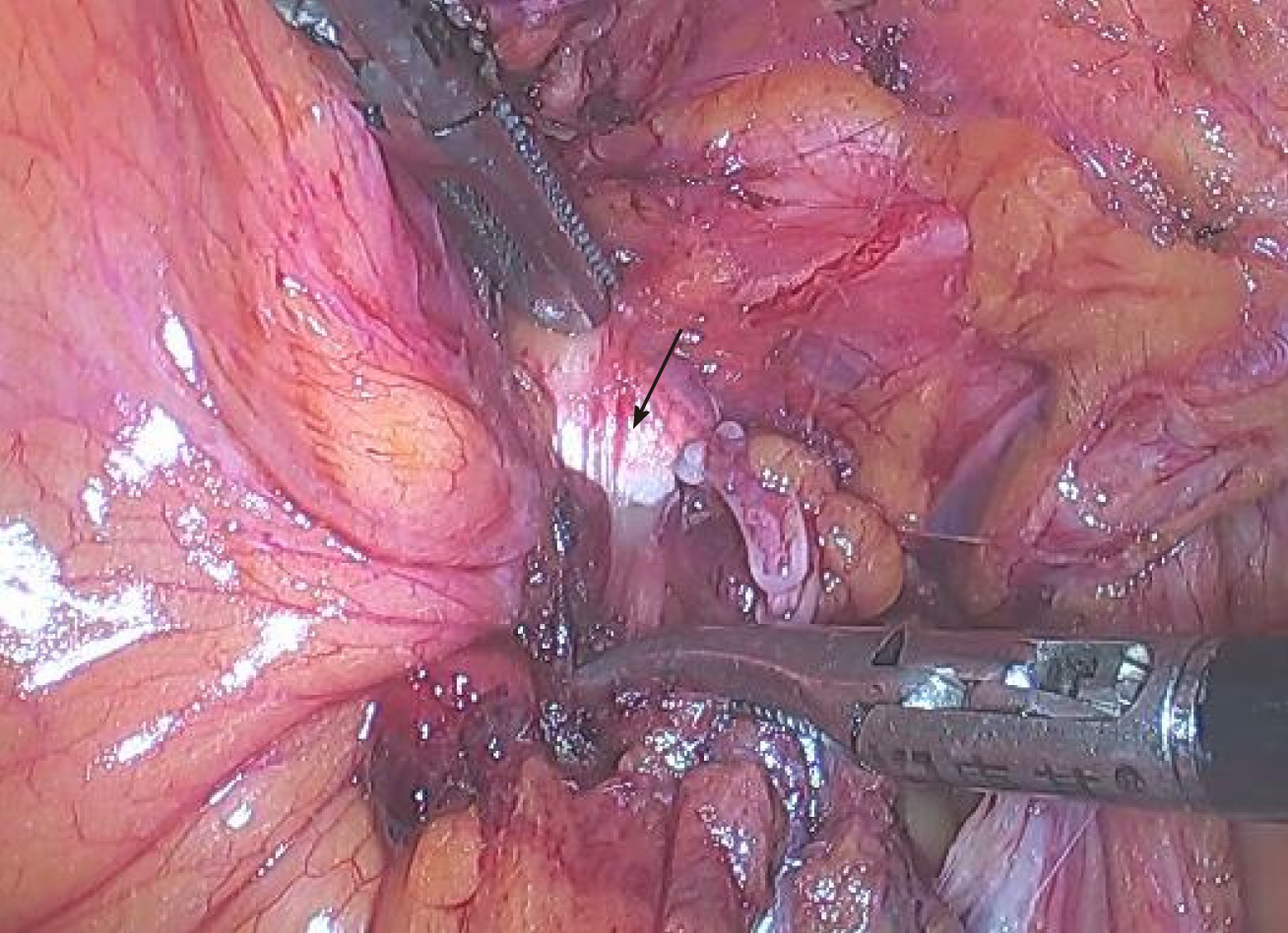
Figure 3 Iliopubic tract.
The arrow shows the iliopubic tract. Part of the iliopubic tract was cut using an electric hook to return the appendix.
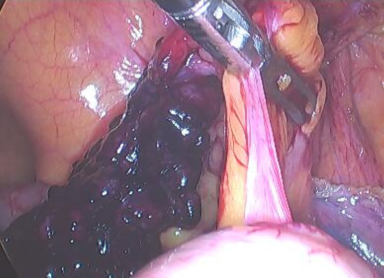
Figure 4 Distal segment of the appendix.
The distal segment of the appendix and its mesangium were dark in color with avascular necrosis and surface exudation.
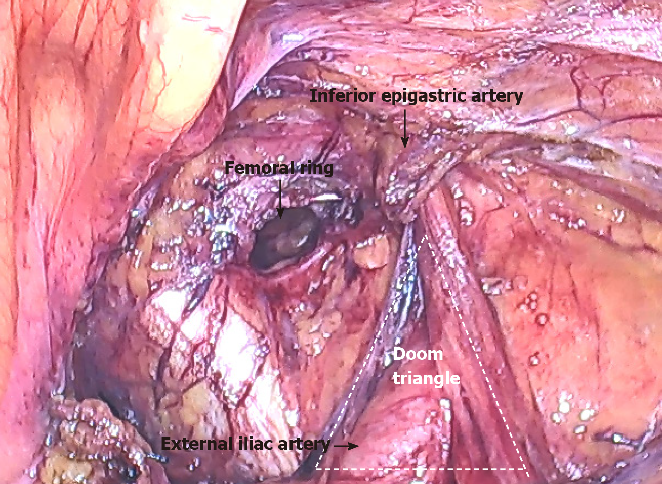
Figure 5 Femoral ring.
After reduction, the femoral ring could be observed. The partially severed iliopubic tract is shown.
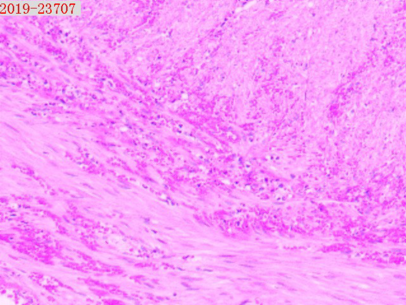
Figure 6 Pathological analysis of the appendix.
The postoperative pathology report showed that a large number of neutrophils had infiltrated the wall of the appendix and mesangial fat, and hemorrhagic necrosis was also observed in the mesangium. Magnification: 50 ×.
- Citation: Yao MQ, Yi BH, Yang Y, Weng XQ, Fan JX, Jiang YP. De Garengeot hernia with avascular necrosis of the appendix: A case report. World J Clin Cases 2021; 9(36): 11355-11361
- URL: https://www.wjgnet.com/2307-8960/full/v9/i36/11355.htm
- DOI: https://dx.doi.org/10.12998/wjcc.v9.i36.11355


