Copyright
©The Author(s) 2021.
World J Clin Cases. Dec 26, 2021; 9(36): 11338-11345
Published online Dec 26, 2021. doi: 10.12998/wjcc.v9.i36.11338
Published online Dec 26, 2021. doi: 10.12998/wjcc.v9.i36.11338
Figure 1 Clinical picture of oral mucormycosis.
A: Multiple lesion on gingiva (Case 1); B: Erythema and multiple pus discharging sinuses on gingiva (Case 2); C: Discoloration of hard palate (Case 4).
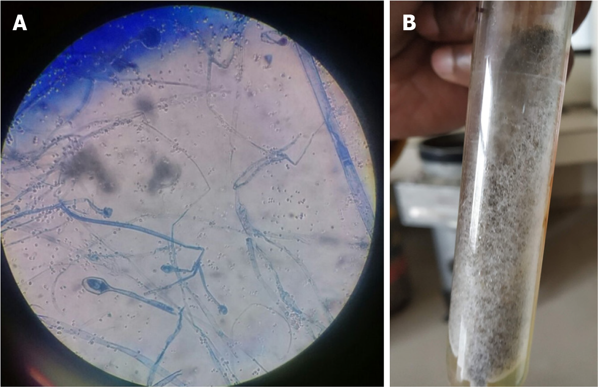
Figure 2 Staining and culture characteristics of mucormycosis.
A: Broad ribbon like aseptate hyphae seen on lactophenol cotton blue staining; B: Whitish grey cottony growth on Sabouraud’s dextrose agar.
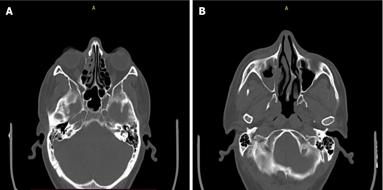
Figure 3 Noncontrast computed tomography of paranasal sinuses.
A: Axial section showing mucosal thickening and bone remodeling involving ethmoidal sinus on right side; B: Axial section showing mucosal thickening on right maxillary sinus with remodeling of medial wall.
- Citation: Upadhyay S, Bharara T, Khandait M, Chawdhry A, Sharma BB. Mucormycosis – resurgence of a deadly opportunist during COVID-19 pandemic: Four case reports. World J Clin Cases 2021; 9(36): 11338-11345
- URL: https://www.wjgnet.com/2307-8960/full/v9/i36/11338.htm
- DOI: https://dx.doi.org/10.12998/wjcc.v9.i36.11338


