Copyright
©The Author(s) 2021.
World J Clin Cases. Dec 16, 2021; 9(35): 11085-11094
Published online Dec 16, 2021. doi: 10.12998/wjcc.v9.i35.11085
Published online Dec 16, 2021. doi: 10.12998/wjcc.v9.i35.11085
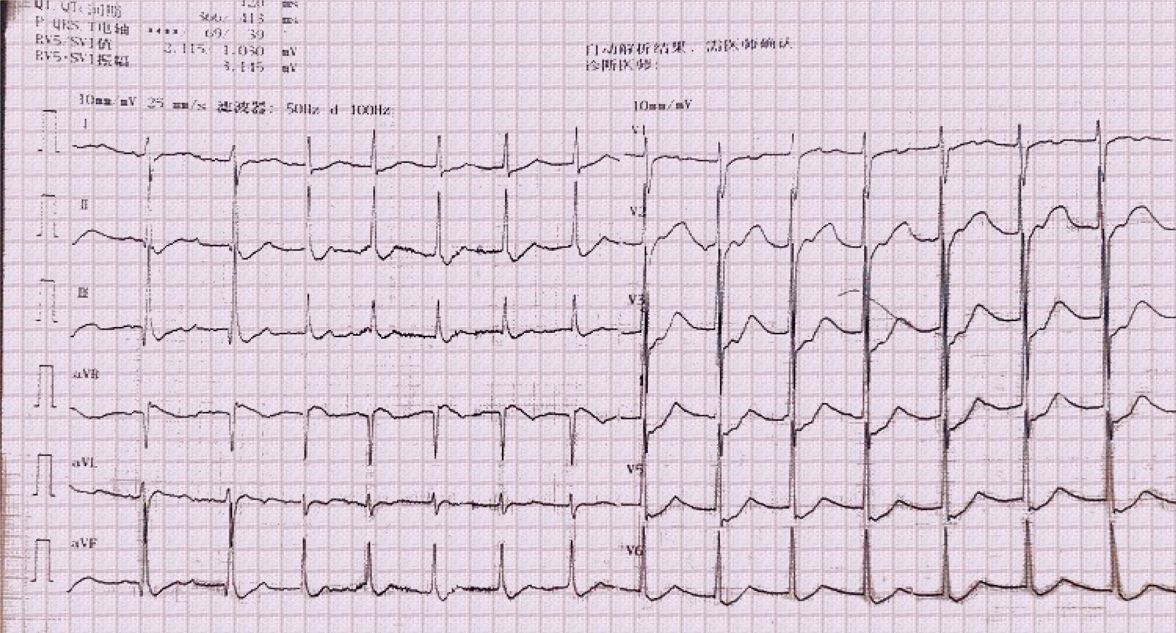
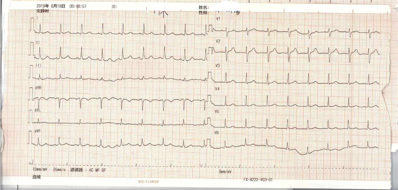
Figure 1 Electrocardiography showed an accelerated junctional rhythm.
No sinus P waves were found, the heart rate was 91 beats per minute, within 60-100 beats per minute, and ST-segment depression was seen in all leads. Accelerated junctional rhythm could be seen in patients with acute myocarditis.
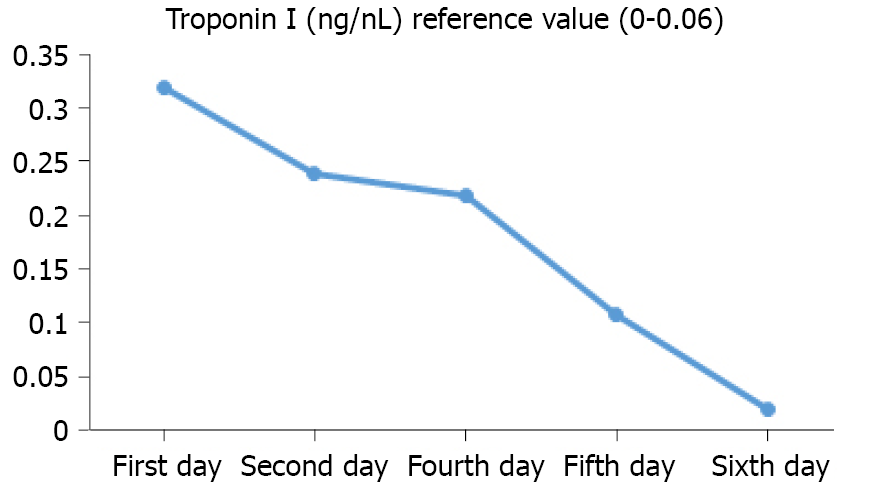
Figure 2 Troponin I level.
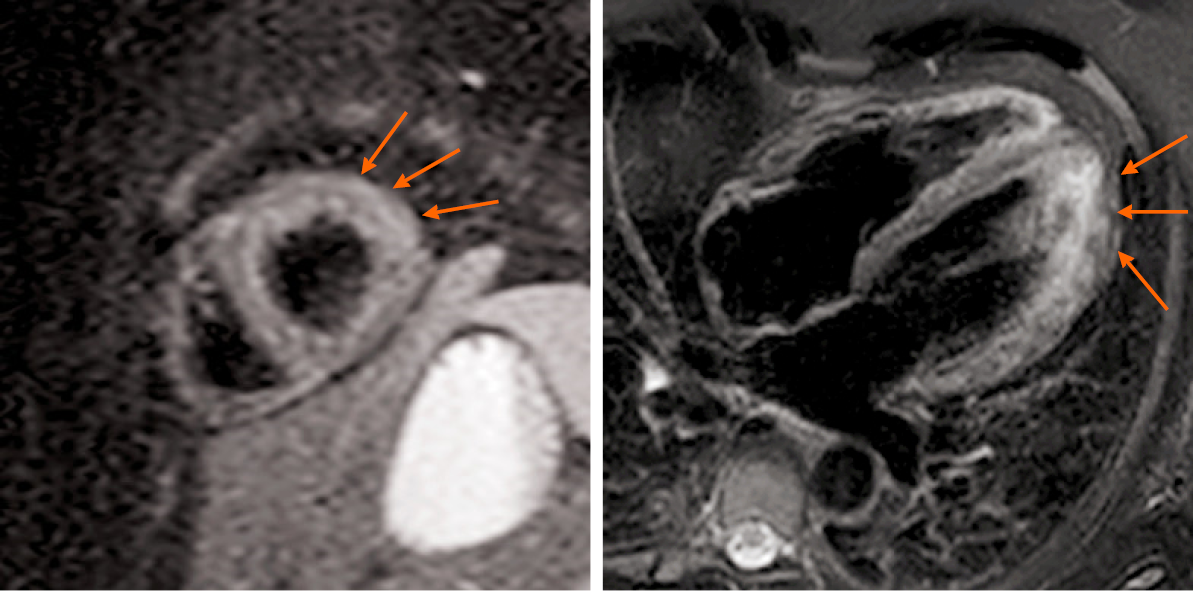
Figure 3 Cardiac magnetic resonance showed myocardial edema in the lateral wall.
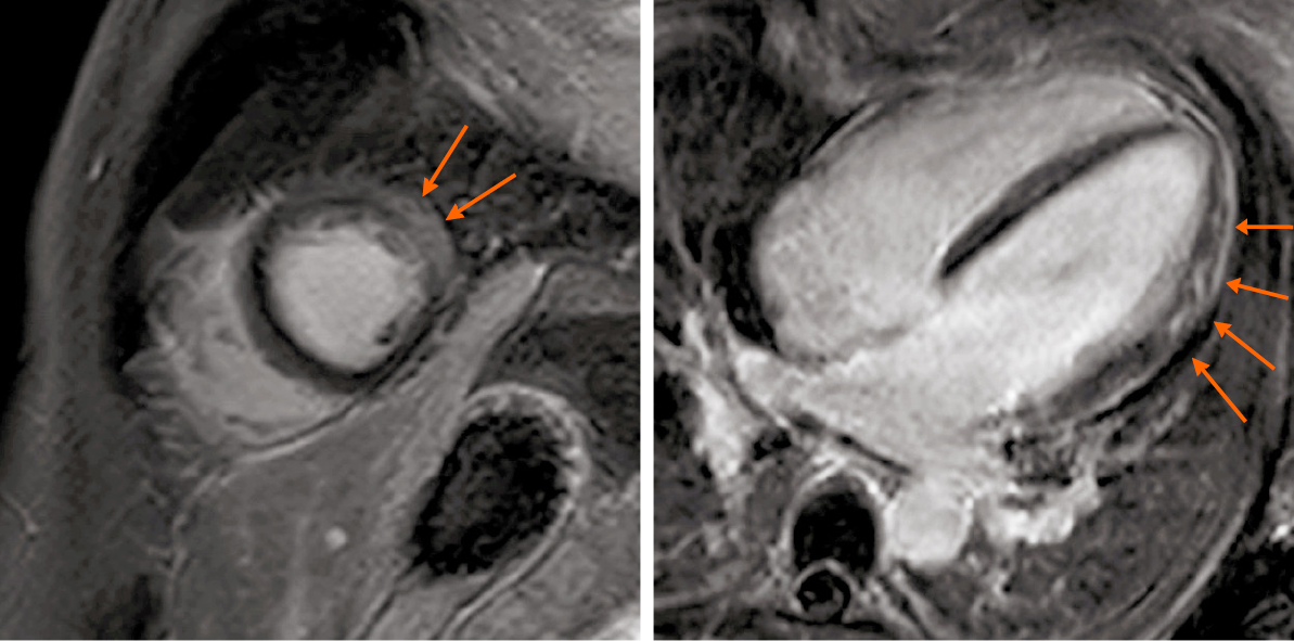
Figure 4 Cardiac magnetic resonance showed intramyocardial and subepicardial late gadolinium enhancement in the lateral apex, anterolateral, and inferior lateral segments of the ventricle.
Figure 5 Sinus rhythm of the patient.
- Citation: Li MM, Liu WS, Shan RC, Teng J, Wang Y. Acute myocarditis presenting as accelerated junctional rhythm in Graves’ disease: A case report. World J Clin Cases 2021; 9(35): 11085-11094
- URL: https://www.wjgnet.com/2307-8960/full/v9/i35/11085.htm
- DOI: https://dx.doi.org/10.12998/wjcc.v9.i35.11085


