Copyright
©The Author(s) 2021.
World J Clin Cases. Dec 16, 2021; 9(35): 11056-11060
Published online Dec 16, 2021. doi: 10.12998/wjcc.v9.i35.11056
Published online Dec 16, 2021. doi: 10.12998/wjcc.v9.i35.11056
Figure 1 Preoperative examination of the mass.
A: Ultrasound; B: Computed tomography; and C: Magnetic resonance imaging.
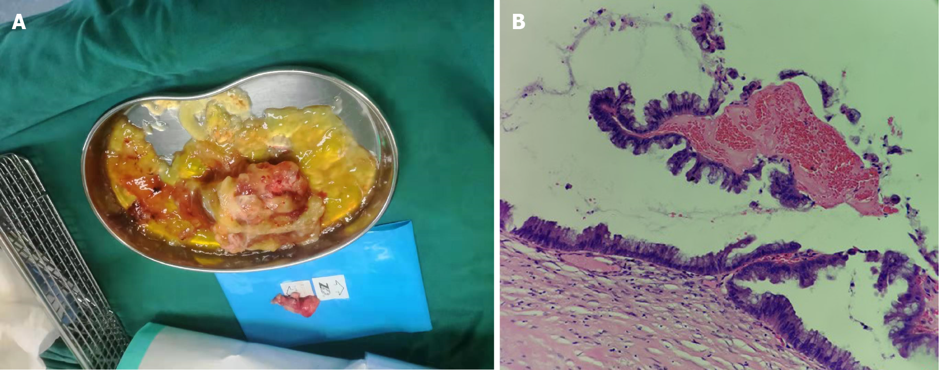
Figure 2 Postoperative images.
A: The macroscopic view the resected mass; and B: Microscopic pathology of the low-grade appendiceal mucinous neoplasm (Magnification × 4).
- Citation: Xu R, Yang ZL. Treatment of a giant low-grade appendiceal mucinous neoplasm: A case report. World J Clin Cases 2021; 9(35): 11056-11060
- URL: https://www.wjgnet.com/2307-8960/full/v9/i35/11056.htm
- DOI: https://dx.doi.org/10.12998/wjcc.v9.i35.11056


