Copyright
©The Author(s) 2021.
World J Clin Cases. Dec 16, 2021; 9(35): 11043-11049
Published online Dec 16, 2021. doi: 10.12998/wjcc.v9.i35.11043
Published online Dec 16, 2021. doi: 10.12998/wjcc.v9.i35.11043
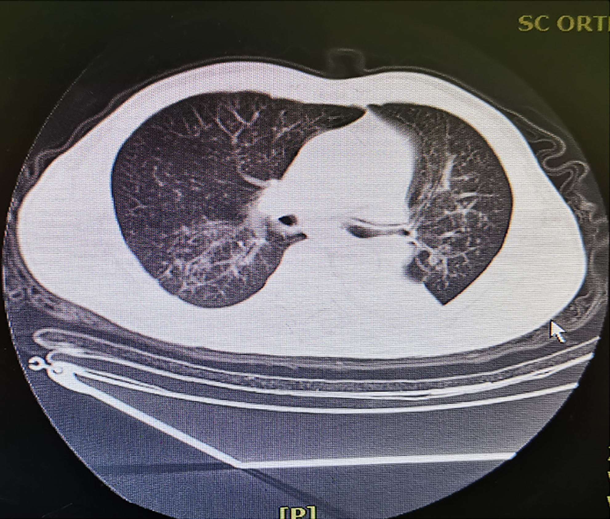
Figure 1 Preoperative computed tomography showing the absence of hemothorax or pneumothorax.
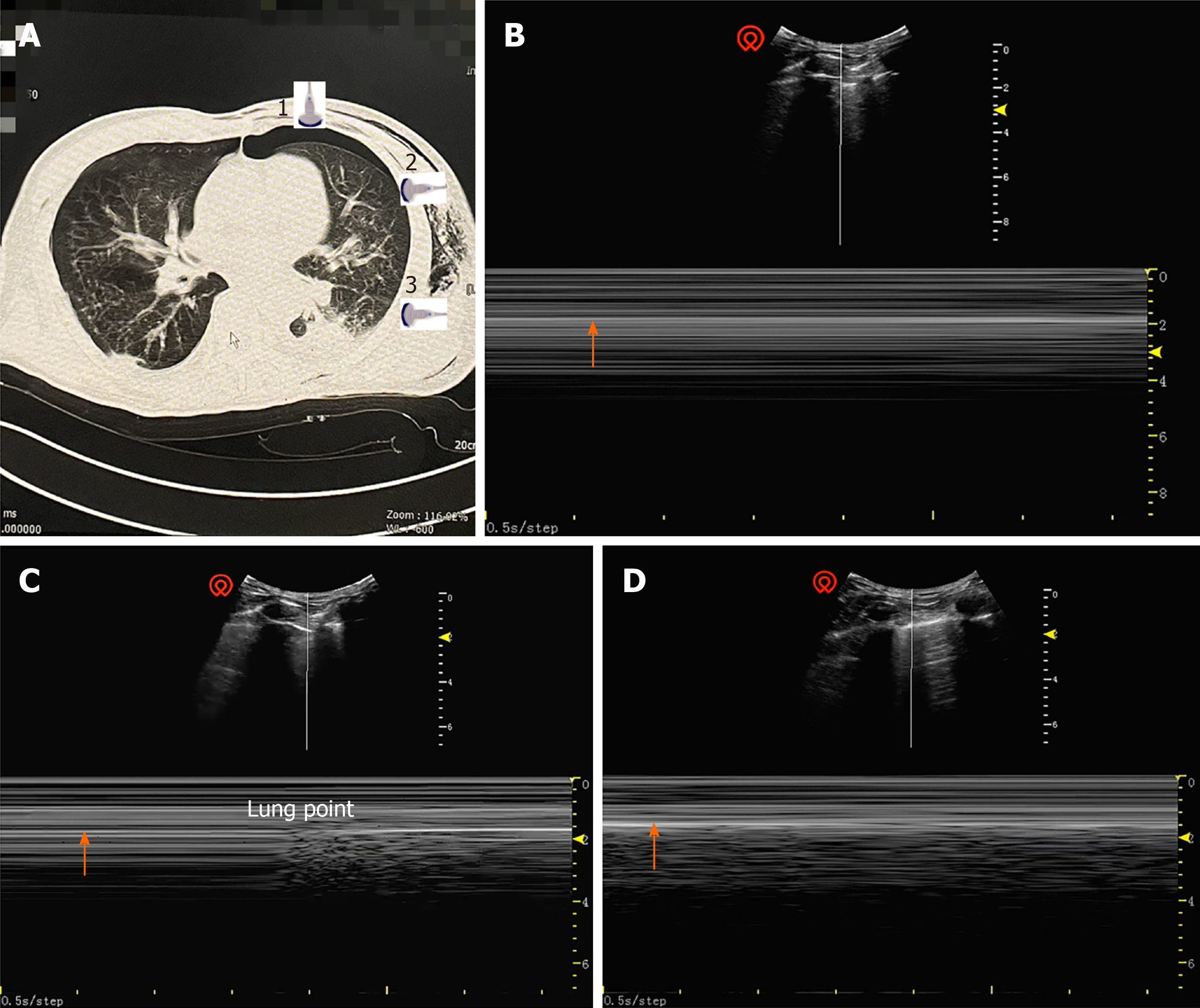
Figure 2 Chest computed tomography and ultrasound.
A: Computed tomography of pneumothorax. Position 1: Pneumothorax; Position 2: Boundary of pneumothorax (lung point); Position 3: No pneumothorax; B: Parallel lines sign with the sonography probe at position 1; C: Lung point sign with the sonography probe at position 2; D: Beach sign with the sonography probe at position 3. White arrows indicate the pleura.
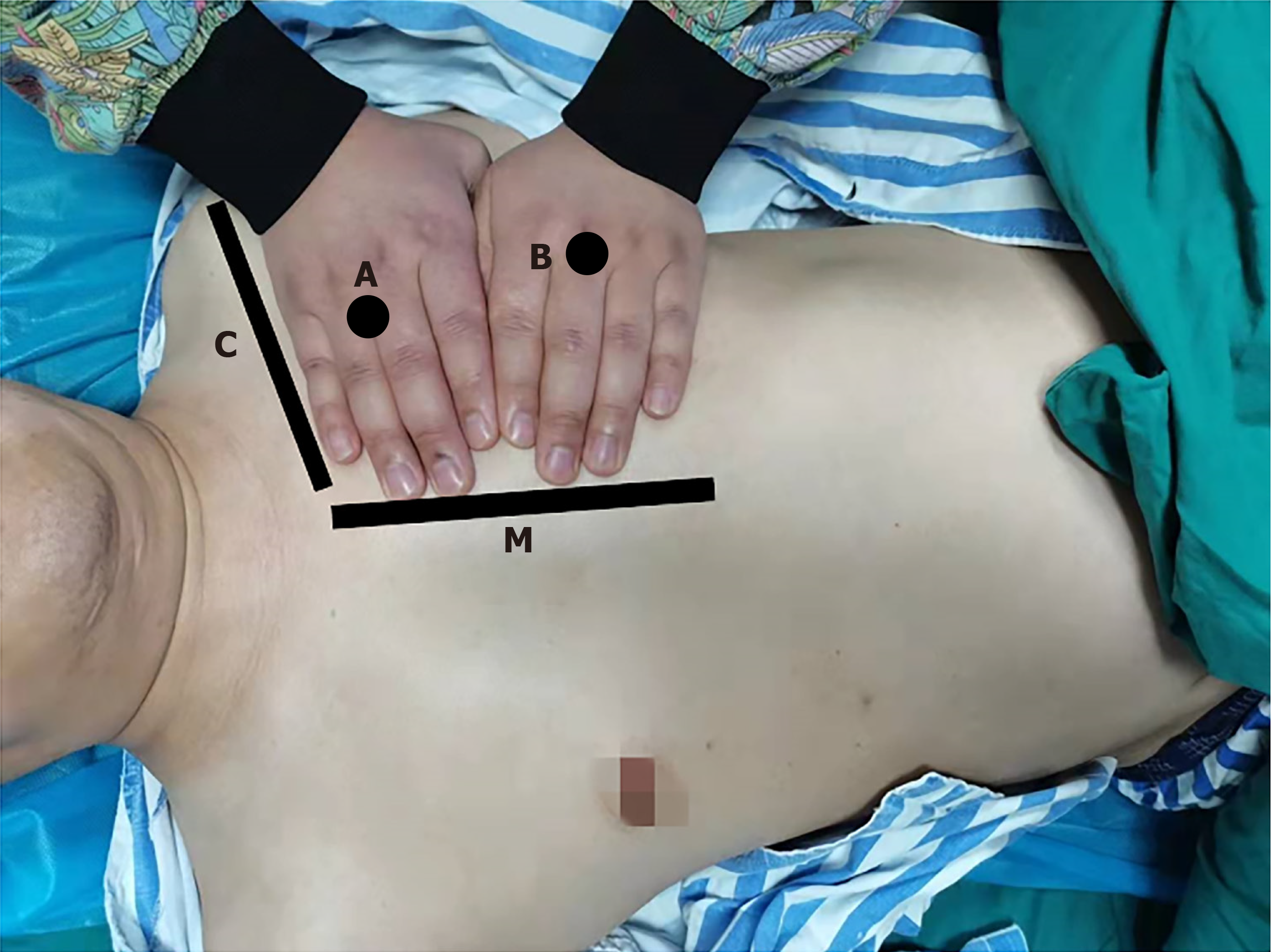
Figure 3 BLUE points recommended for rapid diagnosis or exclusion of perioperative pneumothorax.
A: The upper BLUE point is at the third and fourth metacarpophalangeal joints of the upper hand; B: The lower BLUE point is at the center of the lower palm; C: The black line indicates the clavicle; M: The black line indicates the medial sternal line.
- Citation: Zhang G, Huang XY, Zhang L. Ultrasound guiding the rapid diagnosis and treatment of perioperative pneumothorax: A case report. World J Clin Cases 2021; 9(35): 11043-11049
- URL: https://www.wjgnet.com/2307-8960/full/v9/i35/11043.htm
- DOI: https://dx.doi.org/10.12998/wjcc.v9.i35.11043


