Copyright
©The Author(s) 2021.
World J Clin Cases. Dec 16, 2021; 9(35): 11029-11035
Published online Dec 16, 2021. doi: 10.12998/wjcc.v9.i35.11029
Published online Dec 16, 2021. doi: 10.12998/wjcc.v9.i35.11029
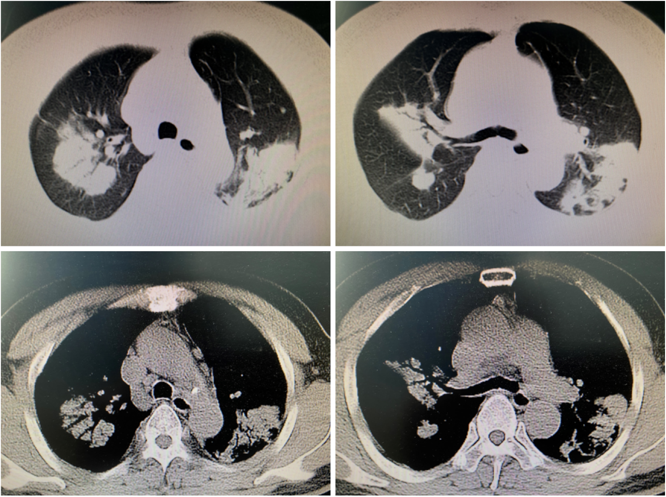
Figure 1 Lung computed tomography showing multiple lung lesions, multiple small nodules with blurred boundaries in both lungs, and cystic changes.
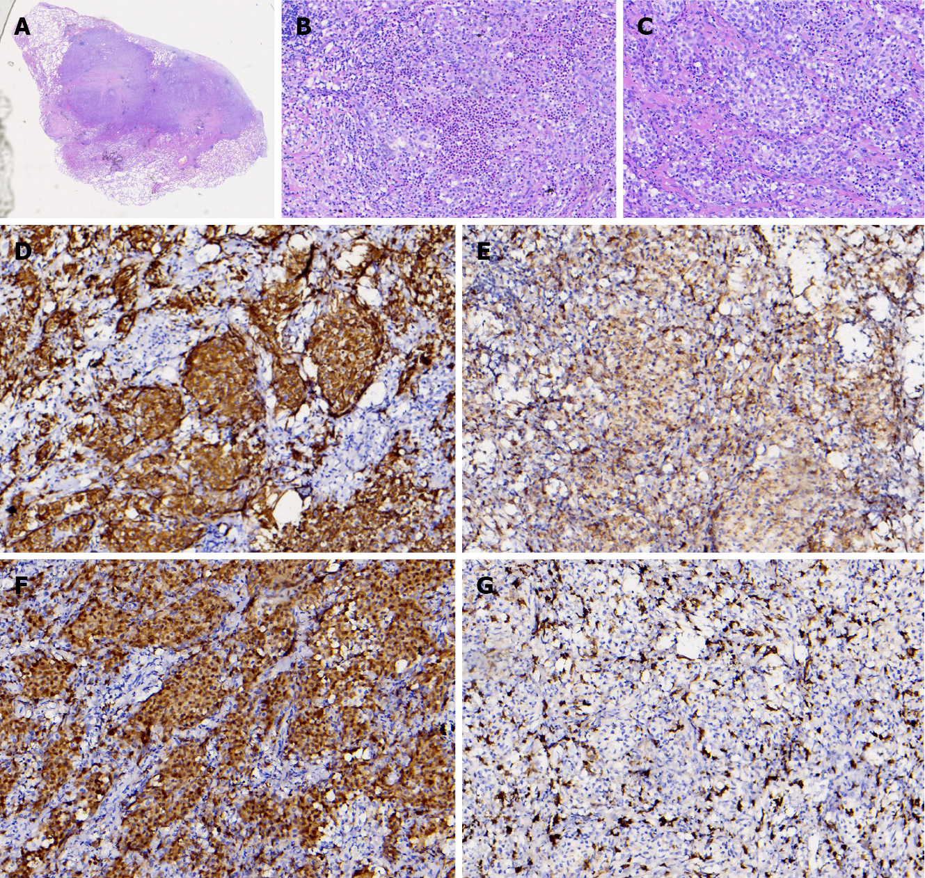
Figure 2 Pathological and immunohistochemistry findings.
A: Lung tissue showing Langerhans infiltrated tissue (magnification: 100 ×); B: Image showing eosinophils with the nucleus stained blue and Langerhans cells (magnification: 200 ×); C: Langerhans cells (magnification: 400 ×); D: Specific immunohistochemical staining for CD1a (+); E: Specific immunohistochemical staining for CD68 (±); F: Specific immunohistochemical staining for S-100 (+); G: Specific immunohistochemical staining for CD163 (-).
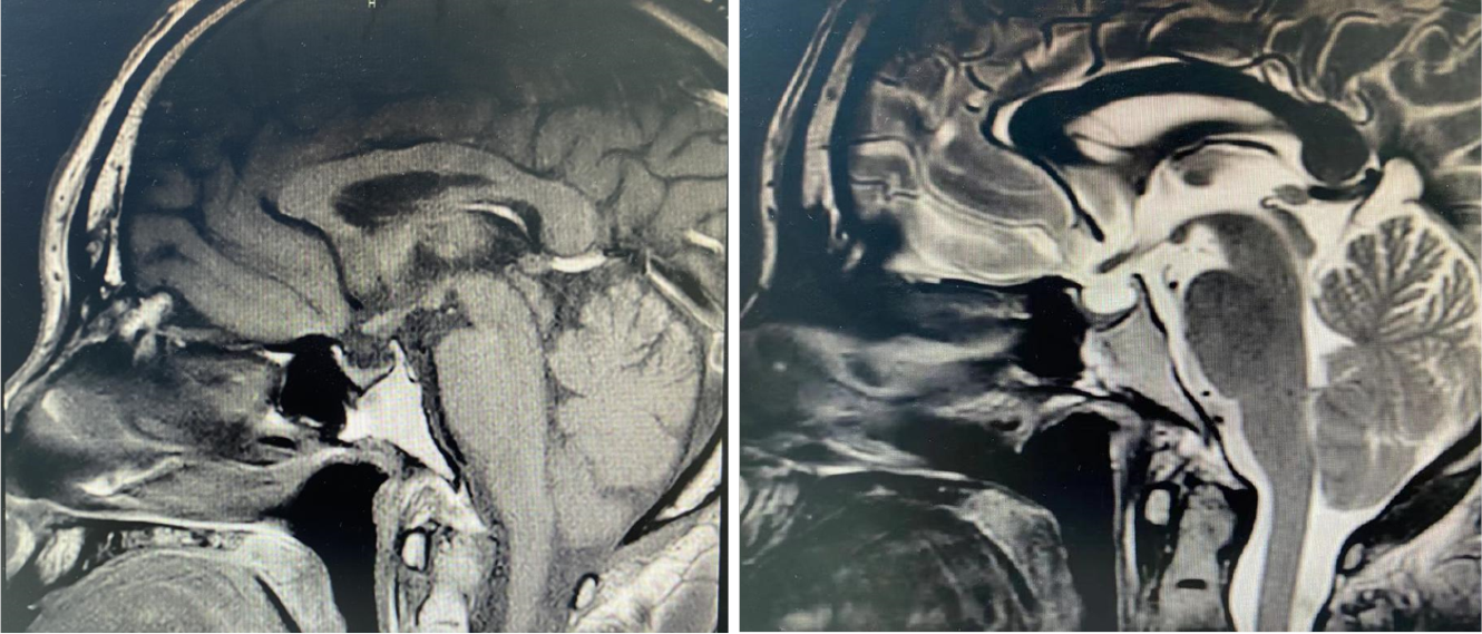
Figure 3 Pituitary magnetic resonance imaging showing vacuolar sella, thinned pituitary stalk, and bilateral maxillary sinus cyst.
- Citation: Luo L, Li YX. Pulmonary Langerhans cell histiocytosis and multiple system involvement: A case report. World J Clin Cases 2021; 9(35): 11029-11035
- URL: https://www.wjgnet.com/2307-8960/full/v9/i35/11029.htm
- DOI: https://dx.doi.org/10.12998/wjcc.v9.i35.11029


