Copyright
©The Author(s) 2021.
World J Clin Cases. Dec 16, 2021; 9(35): 11024-11028
Published online Dec 16, 2021. doi: 10.12998/wjcc.v9.i35.11024
Published online Dec 16, 2021. doi: 10.12998/wjcc.v9.i35.11024
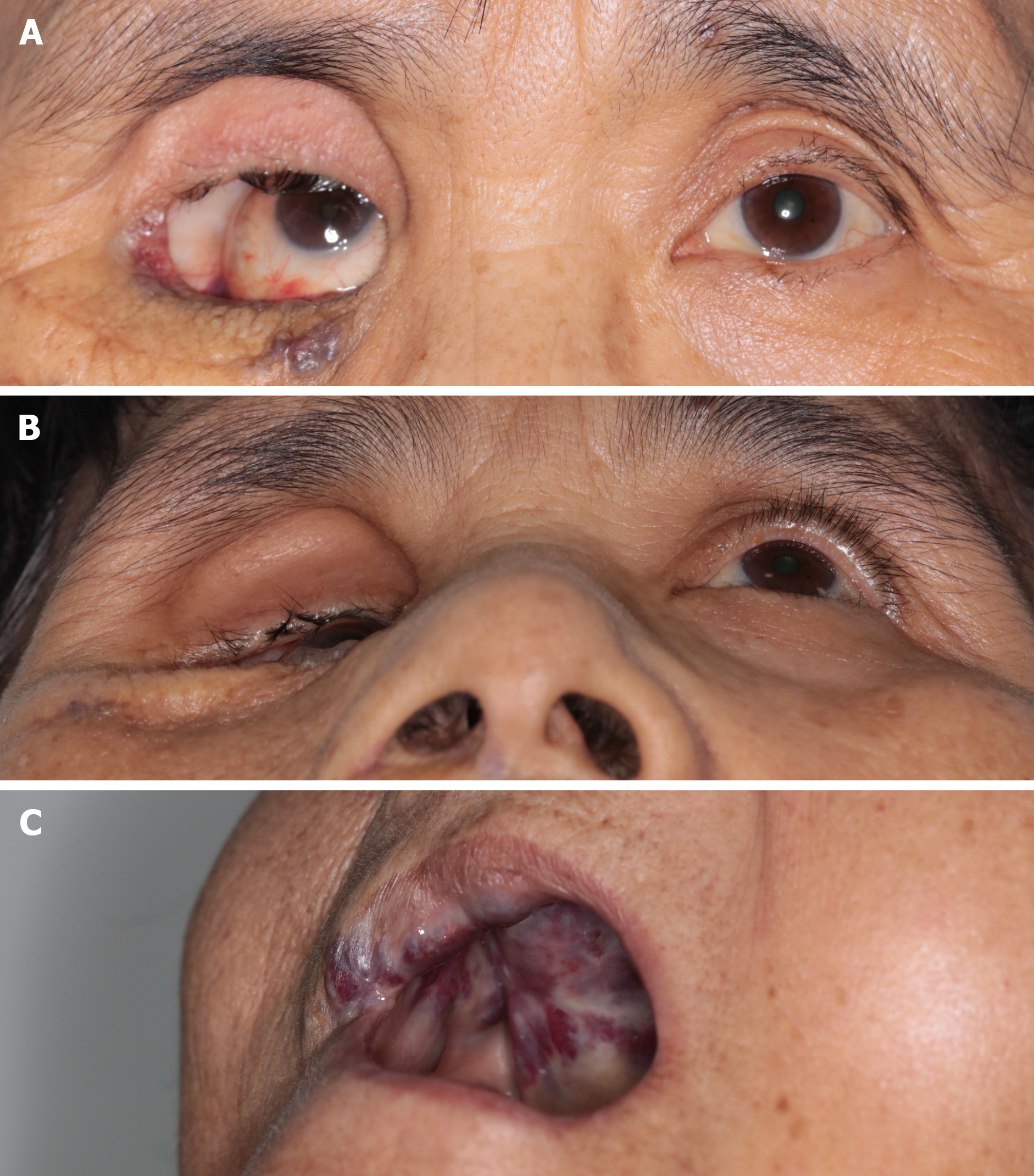
Figure 1 Right eye enophthalmos and entropion.
A: Her right eye was found sunken backwards within the bony orbit, where the orbito-temporal region could be observed; B: Enophthalmos is more obvious when head-up; C: A bulging mass in her hard palate.
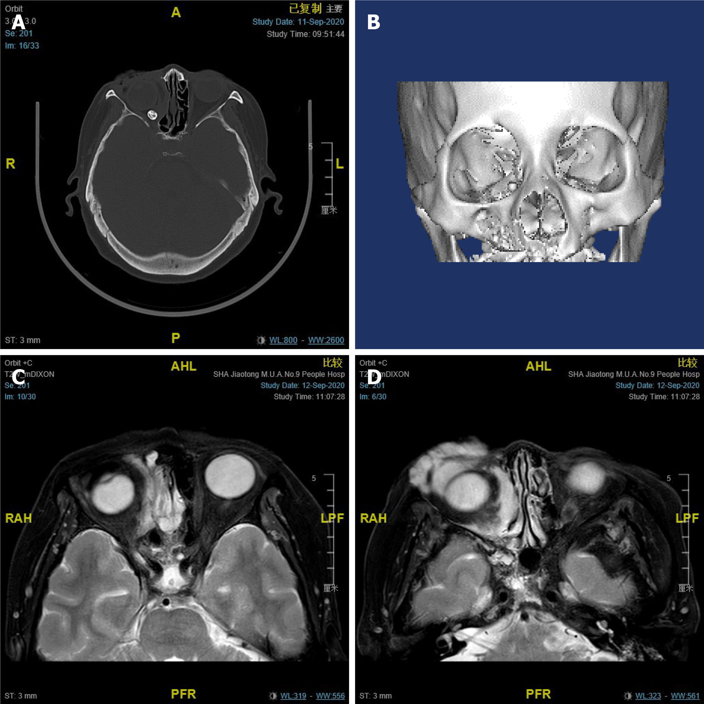
Figure 2 Imaging examinations.
A: The computed tomographic scan showing enophthalmos with local bony defects on the right orbit. A phlebolith is visualized; B: 3D modelling presents an obvious expansion of the right orbital cavity; C and D: Magnetic resonance imaging revealing an absence of orbital soft tissue around her right eye. It also showed an irregular soft tissue mass with indistinct borders inside and outside the muscle pyramid in the right orbit, which could become enlarged after pressurizing.
- Citation: Yang LD, Xu SQ, Wang YF, Jia RB. Severe absence of intra-orbital fat in a patient with orbital venous malformation: A case report. World J Clin Cases 2021; 9(35): 11024-11028
- URL: https://www.wjgnet.com/2307-8960/full/v9/i35/11024.htm
- DOI: https://dx.doi.org/10.12998/wjcc.v9.i35.11024


