Copyright
©The Author(s) 2021.
World J Clin Cases. Dec 16, 2021; 9(35): 10999-11006
Published online Dec 16, 2021. doi: 10.12998/wjcc.v9.i35.10999
Published online Dec 16, 2021. doi: 10.12998/wjcc.v9.i35.10999
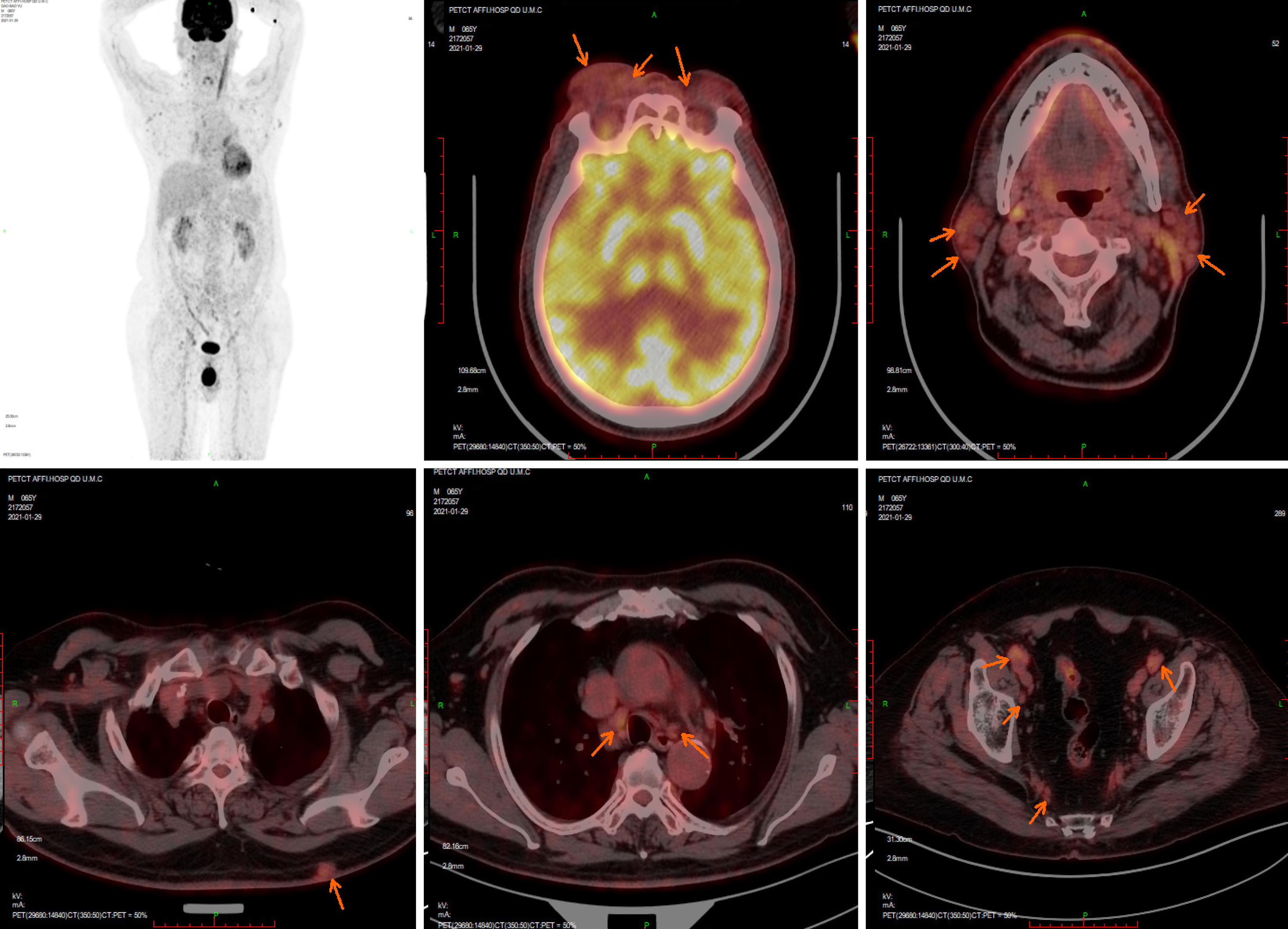
Figure 1 Positron emission tomography computed tomography on January 29, 2020.
Positron emission tomography computed tomography revealed that bilateral ophthalmic muscles, lacrimal glands, intraorbital soft tissue, subcutaneous soft tissue nodules in the back, bilateral mediastinal pleura, and several superficial and deep lymph nodes all showed increased metabolism, accompanied by retroperitoneal fibrosis.
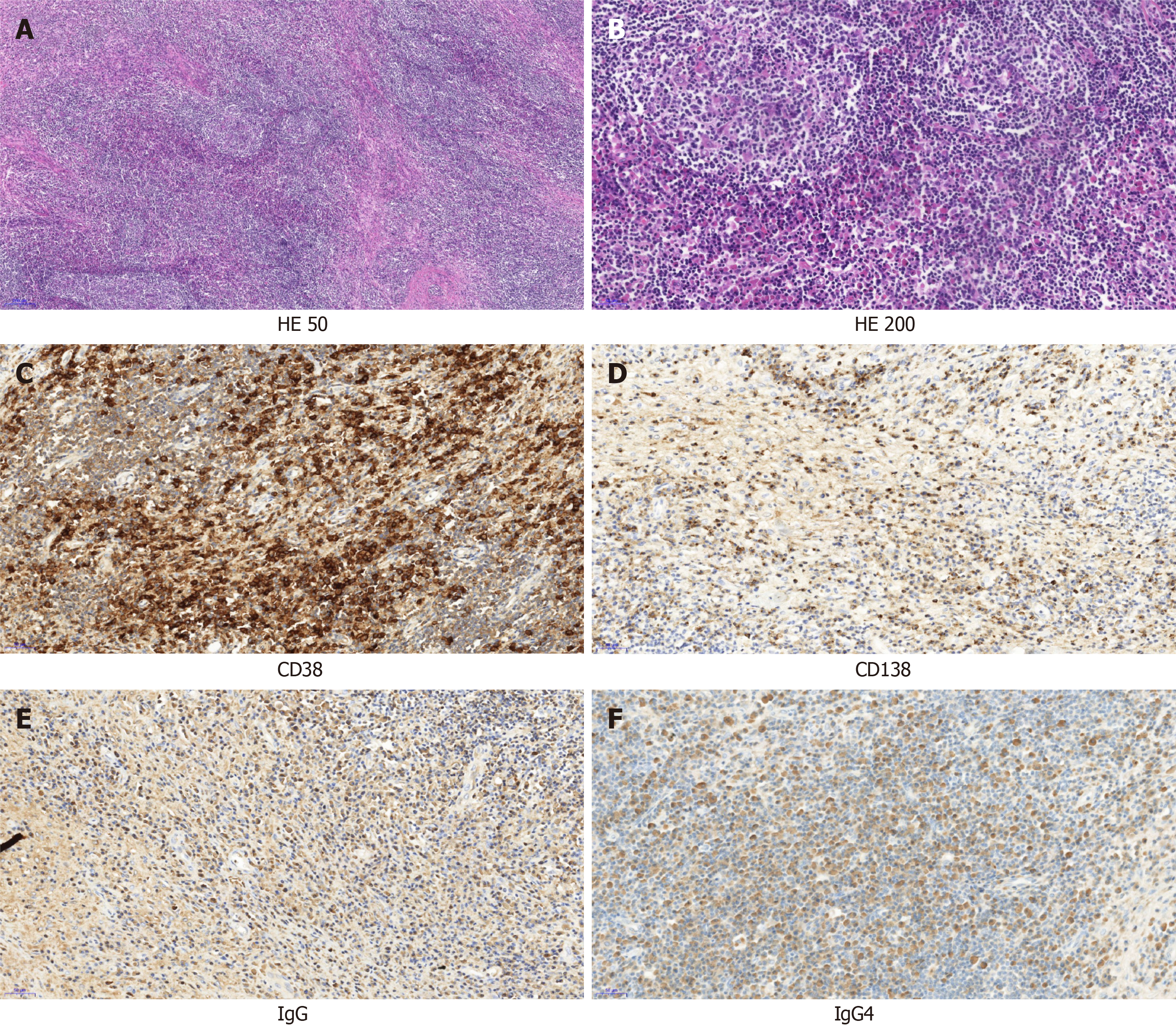
Figure 2 Pathological morphology and immunohistochemistry of orbital mass.
A: At low magnification, the histological morphology showed lymphoproliferative tissue, scattered lymphoproliferative follicles, and interstitial fibrous tissue proliferation (× 50); B: Histological morphology at high magnification showed hyperplasia of small vessels in the follicles, and a large number of plasma cells infiltrated between the follicles (× 200); C: CD38 immunohistochemical staining showed a large number of positive plasma cells in the interfollicular space; D: Immunohistochemical staining of CD138 showed a large number of positive plasma cells in the interfollicle; E: Immunoglobulin (Ig) G positive plasma cells could be seen by immunohistochemical staining; F: IgG4-positive plasma cells could be seen by immunohistochemical staining; The IgG4/IgG ratio was greater than 40%. HE: Hematoxylin and eosin stain; Ig: Immunoglobulin.
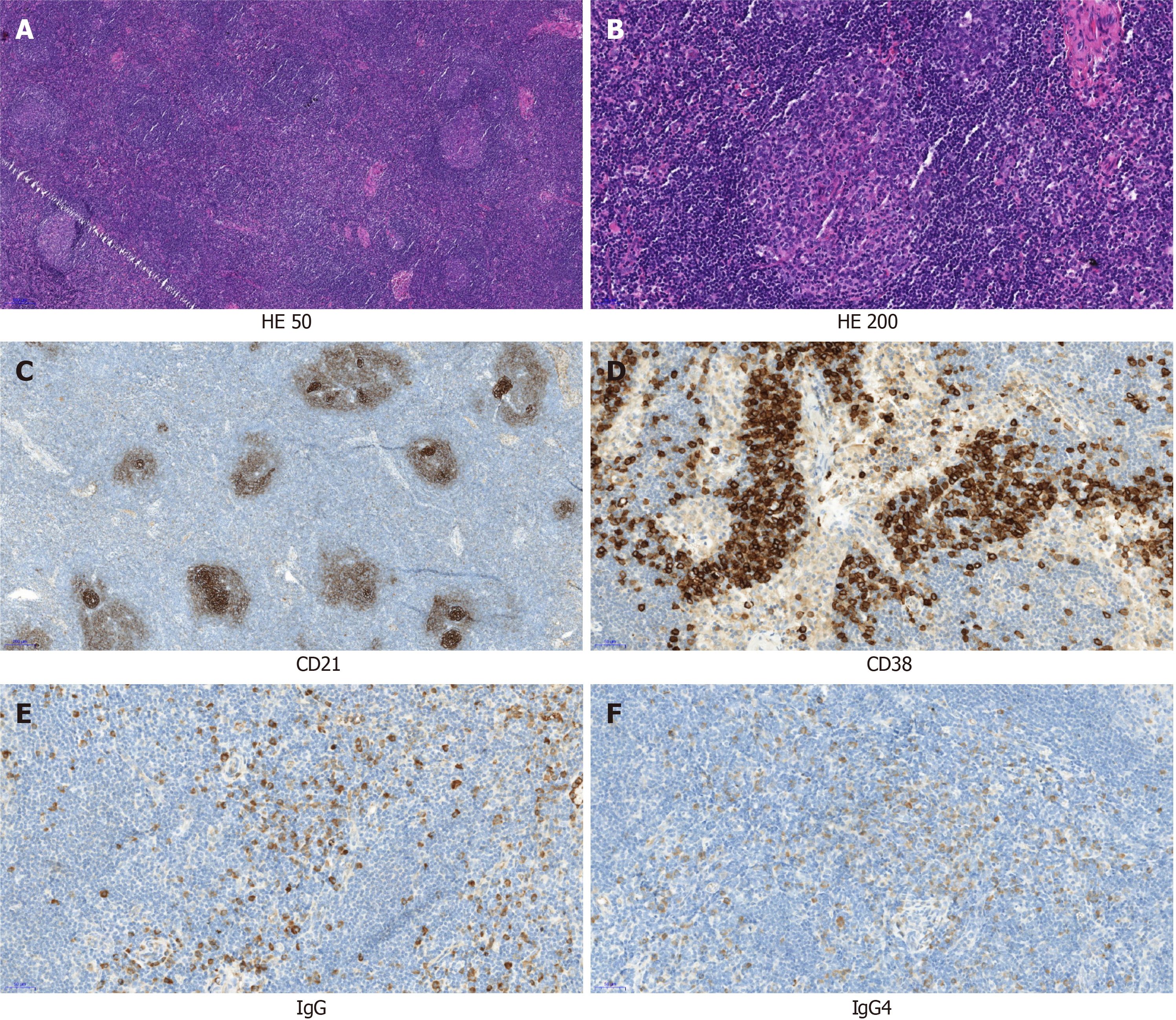
Figure 3 Pathological morphology and immunohistochemistry of cervical lymph nodes.
A: At low power histological morphology, most of the lymphoid sinuses of the lymph nodes disappeared, and proliferative lymphatic follicles were evenly distributed throughout the lymph nodes. Lymphocytes in the mantle region were widened, and small blood vessels between the follicles were increased, with partial hyalinization, similar to Castleman disease morphological changes (× 50); B: At high magnification, small blood vessels in the follicles were observed to grow and proliferate, a large amount of lymphatic tissue proliferated between the follicles, and plasma cells were infiltrated (× 200); C: Immunohistochemical staining of CD21 showed a network of follicular dendritic cells scattered throughout the lymphatic follicles of the lymph node; D: CD38 immunohistochemical staining showed a large number of positive plasma cells in the interfollicular space; E: Immunoglobulin (Ig) G positive plasma cells could be seen by immunohistochemical staining; F: IgG4-positive plasma cells could be seen by immunohistochemical staining; the IgG4/IgG ratio was greater than 40%. HE: Hematoxylin and eosin stain; Ig: Immunoglobulin.
- Citation: Hao FY, Yang FX, Bian HY, Zhao X. Immunoglobulin G4-related lymph node disease with an orbital mass mimicking Castleman disease: A case report. World J Clin Cases 2021; 9(35): 10999-11006
- URL: https://www.wjgnet.com/2307-8960/full/v9/i35/10999.htm
- DOI: https://dx.doi.org/10.12998/wjcc.v9.i35.10999


