Copyright
©The Author(s) 2021.
World J Clin Cases. Dec 16, 2021; 9(35): 10994-10998
Published online Dec 16, 2021. doi: 10.12998/wjcc.v9.i35.10994
Published online Dec 16, 2021. doi: 10.12998/wjcc.v9.i35.10994
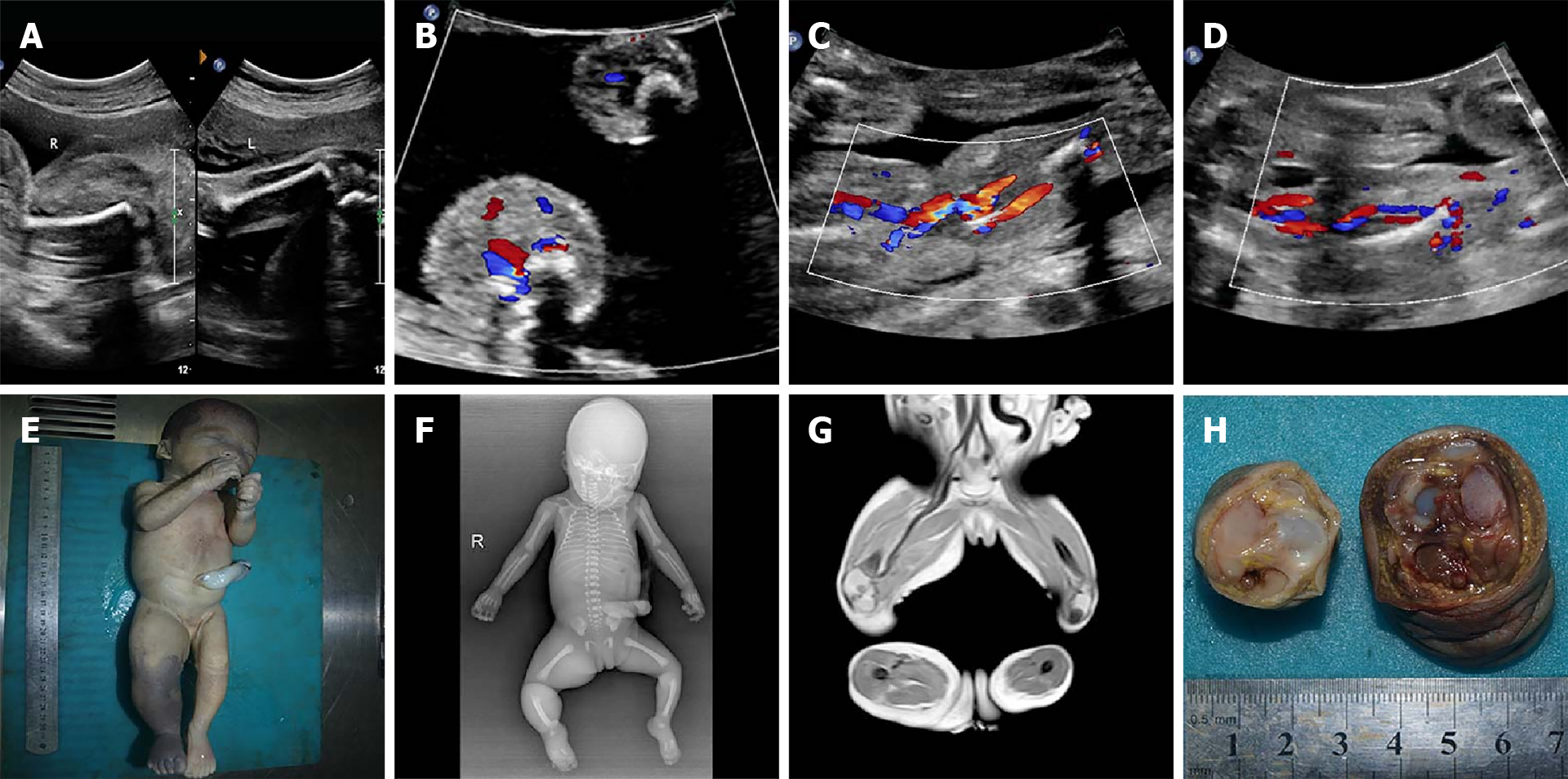
Figure 1 Prenatal sonograms at 18 wk of gestation and images after induced-abortion.
A and B: Marked thickening of the right thigh, no marked edema and fluid/cystic spaces in the lower limbs (A, sagittal section and B, transverse section); C and D: Enlarged vasculature and higher intensive blood flow signals in the right limb; E: The purple stains predominantly affecting the right lower limb and asymmetry of the lower limbs; F: X-ray image showing the right lower limb hypertrophy; G: Magnetic resonance imaging image showing enlargement of the vasculature and hypertrophy of soft tissue; H: Severe congestion in the right lower limb. R: Right lower limb; L: Left lower limb.
- Citation: Pang HQ, Gao QQ. Prenatal ultrasonographic findings in Klippel-Trenaunay syndrome: A case report. World J Clin Cases 2021; 9(35): 10994-10998
- URL: https://www.wjgnet.com/2307-8960/full/v9/i35/10994.htm
- DOI: https://dx.doi.org/10.12998/wjcc.v9.i35.10994


