Copyright
©The Author(s) 2021.
World J Clin Cases. Dec 6, 2021; 9(34): 10696-10701
Published online Dec 6, 2021. doi: 10.12998/wjcc.v9.i34.10696
Published online Dec 6, 2021. doi: 10.12998/wjcc.v9.i34.10696
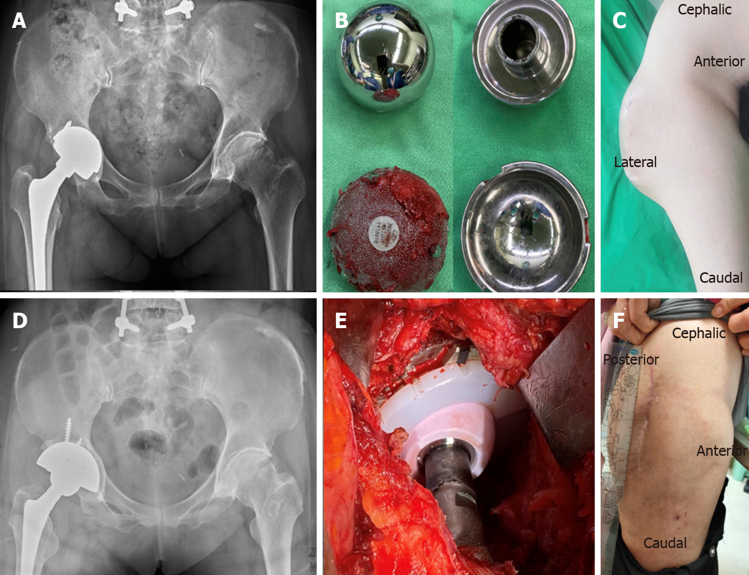
Figure 1 Pre-operative and post-operative series images.
A: Radiography of metal-on-metal total hip arthroplasty (THA) over right hip; B: The metal-on-metal acetabular cup and femoral head removed from revision surgery. Surface scratching and erosion was observed between the cup/head component; C: Pre-operative gross photo (AP view) of right hip showed mass protruding at lateral side of right hip; D and E: Radiography and gross photo of revision ceramic-on-polyethylene THA; F: Post-revision gross photo (lateral view) of patient after locally tigecycline injection showed subsidence of the mass at 24-mo follow-up.
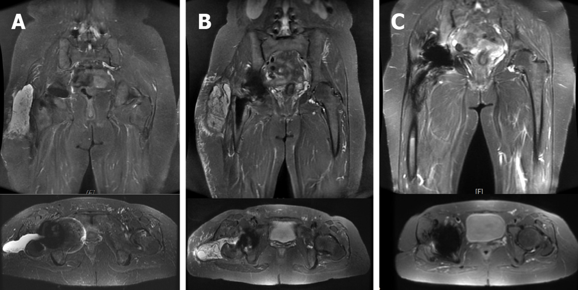
Figure 2 Magnetic resonance imaging series of pre-operative, post-revision surgery, and after Tigecycline local treatment.
A: Initial radiographic evaluation: magnetic resonance imaging (MRI) image of right hip showed joint effusion and trochanteric bursitis involving right lateral subcutaneous layer of right artificial hip. The finding corresponds to the diagnosis of pseudotumor; B: MRI image following revision total hip arthroplasty (THA): recurrent periprosthetic pseudotumor over right hip to lateral subcutaneous layer and adjacent subcutaneous inflammation; C: MRI image following revision THA, one month after local tigecycline infusion, showed subsidence of recurrent pseudotumor, periprosthetic soft tissue swelling and effusion.
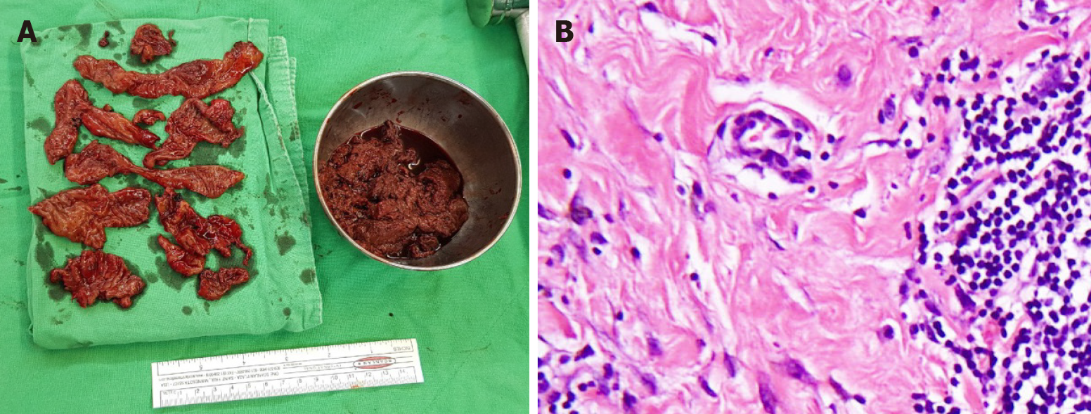
Figure 3 Gross photo and pathological section of resected pseudotumor.
A: Photograph of debrided necrotic tissues taken from arthrotomy and removal of pseudotumor; B: Synovial lining cell hyperplasia with lymphocytic cells infiltration and Stromal fibroplasia, with the histological diagnosis of aseptic aseptic lymphocyte-dominant vasculitis-associated lesion.
- Citation: Lin IH, Tsai CH. Tigecycline sclerotherapy for recurrent pseudotumor in aseptic lymphocyte-dominant vasculitis-associated lesion after metal-on-metal total hip arthroplasty: A case report. World J Clin Cases 2021; 9(34): 10696-10701
- URL: https://www.wjgnet.com/2307-8960/full/v9/i34/10696.htm
- DOI: https://dx.doi.org/10.12998/wjcc.v9.i34.10696


