Copyright
©The Author(s) 2021.
World J Clin Cases. Dec 6, 2021; 9(34): 10689-10695
Published online Dec 6, 2021. doi: 10.12998/wjcc.v9.i34.10689
Published online Dec 6, 2021. doi: 10.12998/wjcc.v9.i34.10689
Figure 1 Computed tomography angiography results prior to thoracic endovascular aortic repair.
A: True lumen collapse (arrow); B: The maximal diameter of the distal tear was 10.71 mm; C: The maximal diameter of the descending aorta was 51.8 mm; D: Type III aortic arch, with the dissecting aneurysm being immediately adjacent to the left subclavian artery.
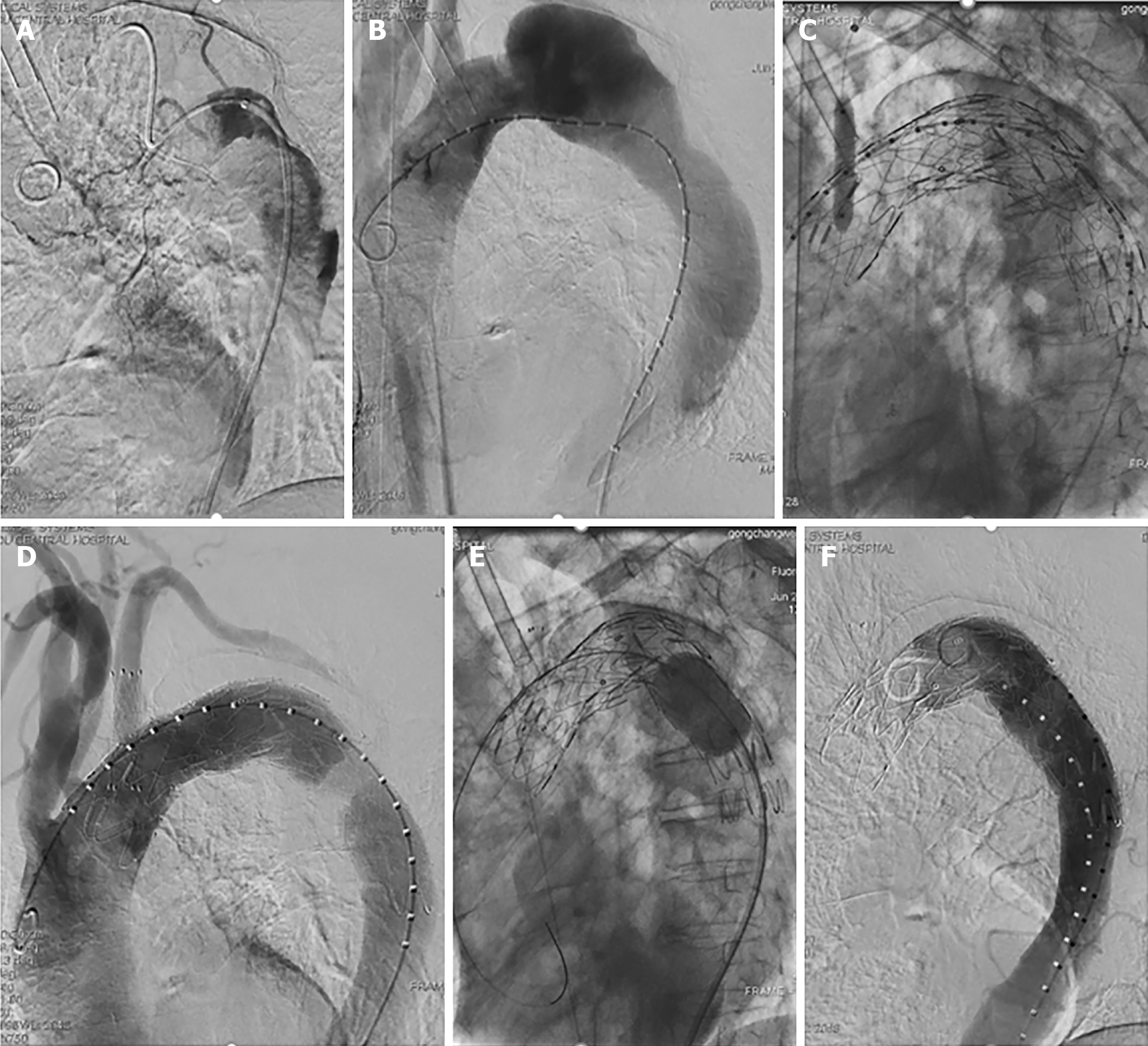
Figure 2 Aortic angiography.
A: The pigtail catheter adopted a helical shape in the true lumen, with the proximal true lumen being clearly compressed; B: The helical shape of the pigtail catheter was corrected, with aortic angiography showing the large false lumen; C: A balloon dilatation catheter was used to enlarge the small hole in the ANKURA stent graft; D: Aortic angiography revealed no evidence of endoleakage and good blood flow in the left subclavian artery; E: The ANKURA stent graft was enlarged with a balloon dilatation catheter; F: The stent shape appeared satisfactory upon angiographic reassessment.
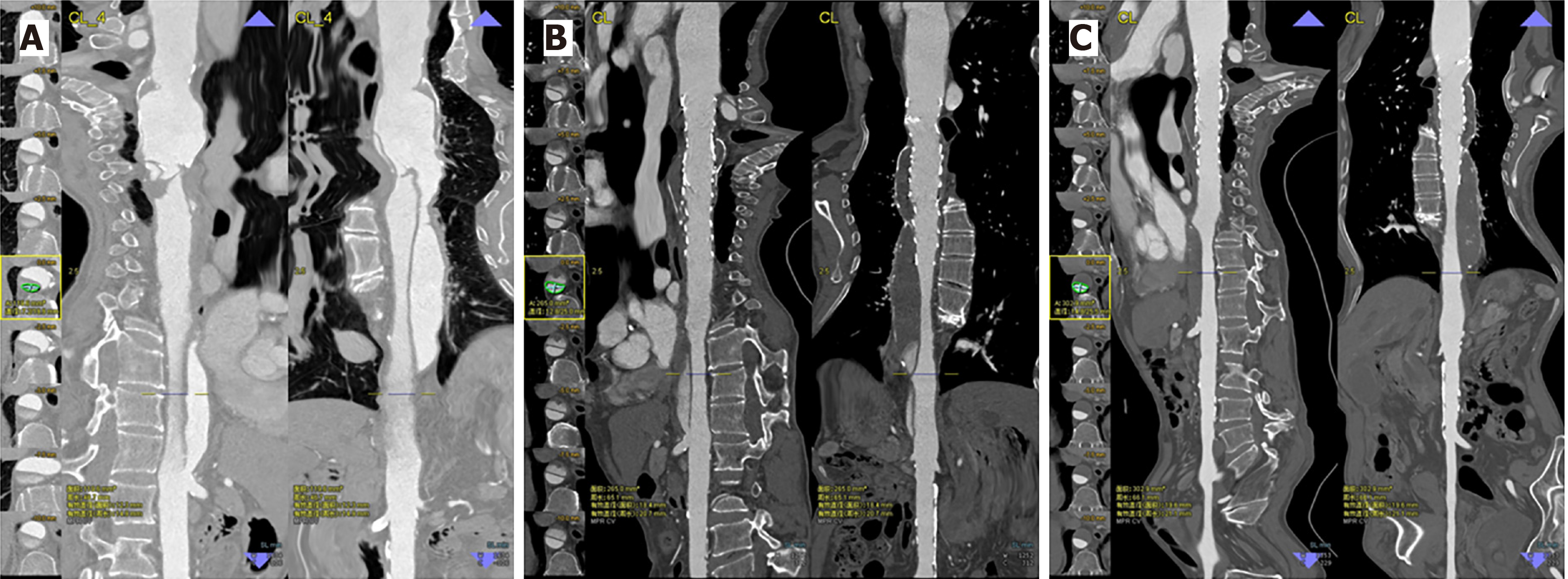
Figure 3 The false lumen gradually decreased in size and became completely thrombotic.
A: Prior to thoracic endovascular aortic repair; B: 3-mo follow-up; C: 28-mo follow-up.
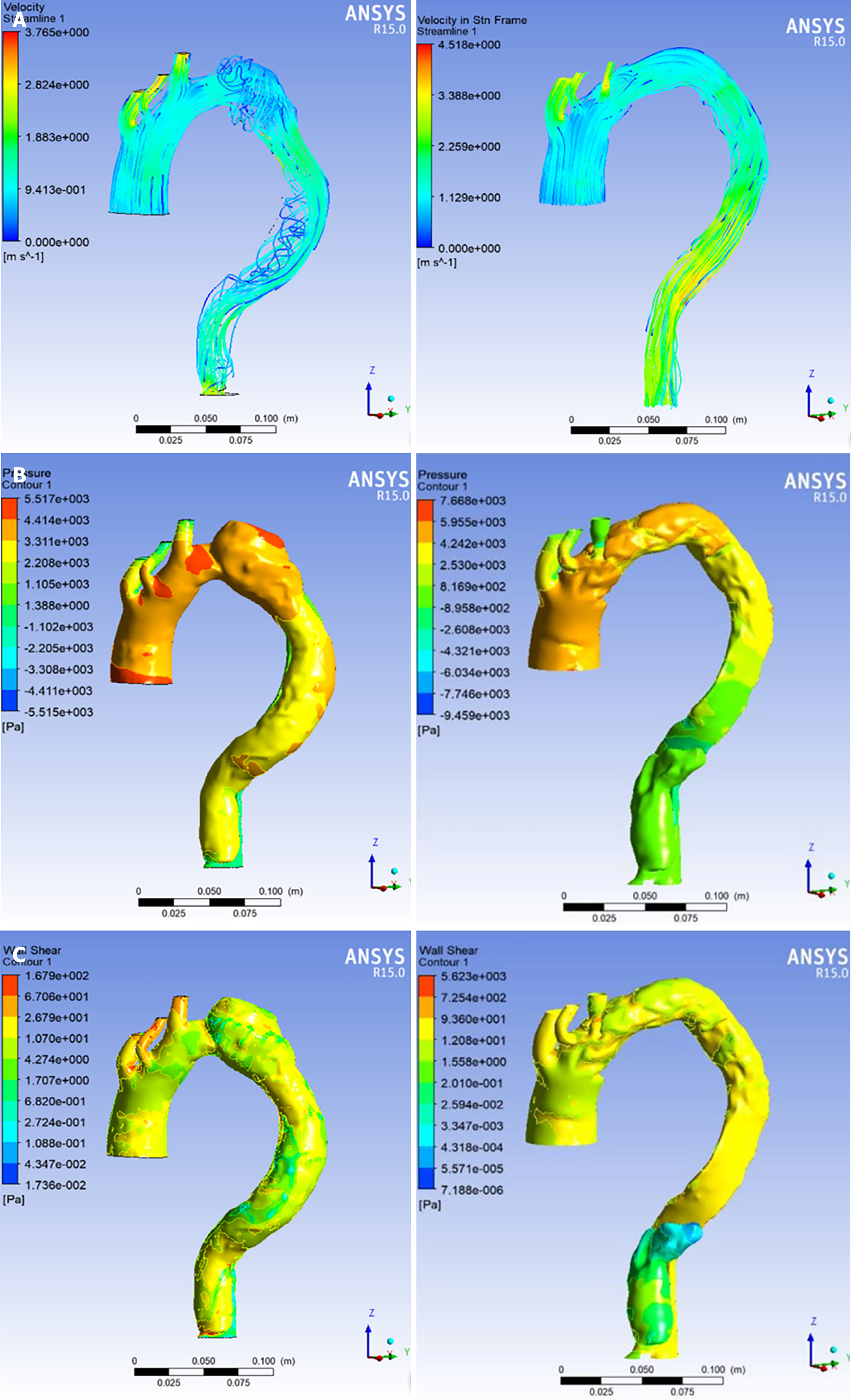
Figure 4 Computational hemodynamic analysis in the true and false lumen before and after thoracic endovascular aortic repair treatment.
A: Blood flow feature; B: Pressure; C: Wall shear.
- Citation: Zhang L, Guan WK, Wu HP, Li X, Lv KP, Zeng CL, Song HH, Ye QL. Proximal true lumen collapse in a chronic type B aortic dissection patient: A case report. World J Clin Cases 2021; 9(34): 10689-10695
- URL: https://www.wjgnet.com/2307-8960/full/v9/i34/10689.htm
- DOI: https://dx.doi.org/10.12998/wjcc.v9.i34.10689


