Copyright
©The Author(s) 2021.
World J Clin Cases. Dec 6, 2021; 9(34): 10671-10680
Published online Dec 6, 2021. doi: 10.12998/wjcc.v9.i34.10671
Published online Dec 6, 2021. doi: 10.12998/wjcc.v9.i34.10671
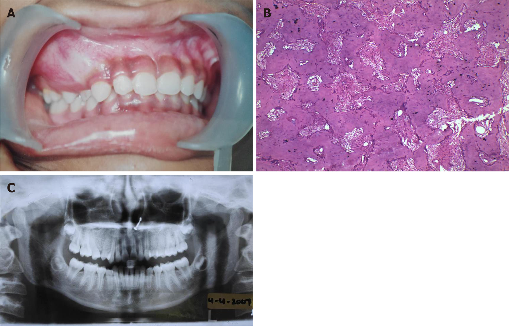
Figure 1 Pre-surgical treatment (April 2009).
A: Intra-oral photograph; B: Immuno-histopathology slide; C: Orthopantomogram X-ray.
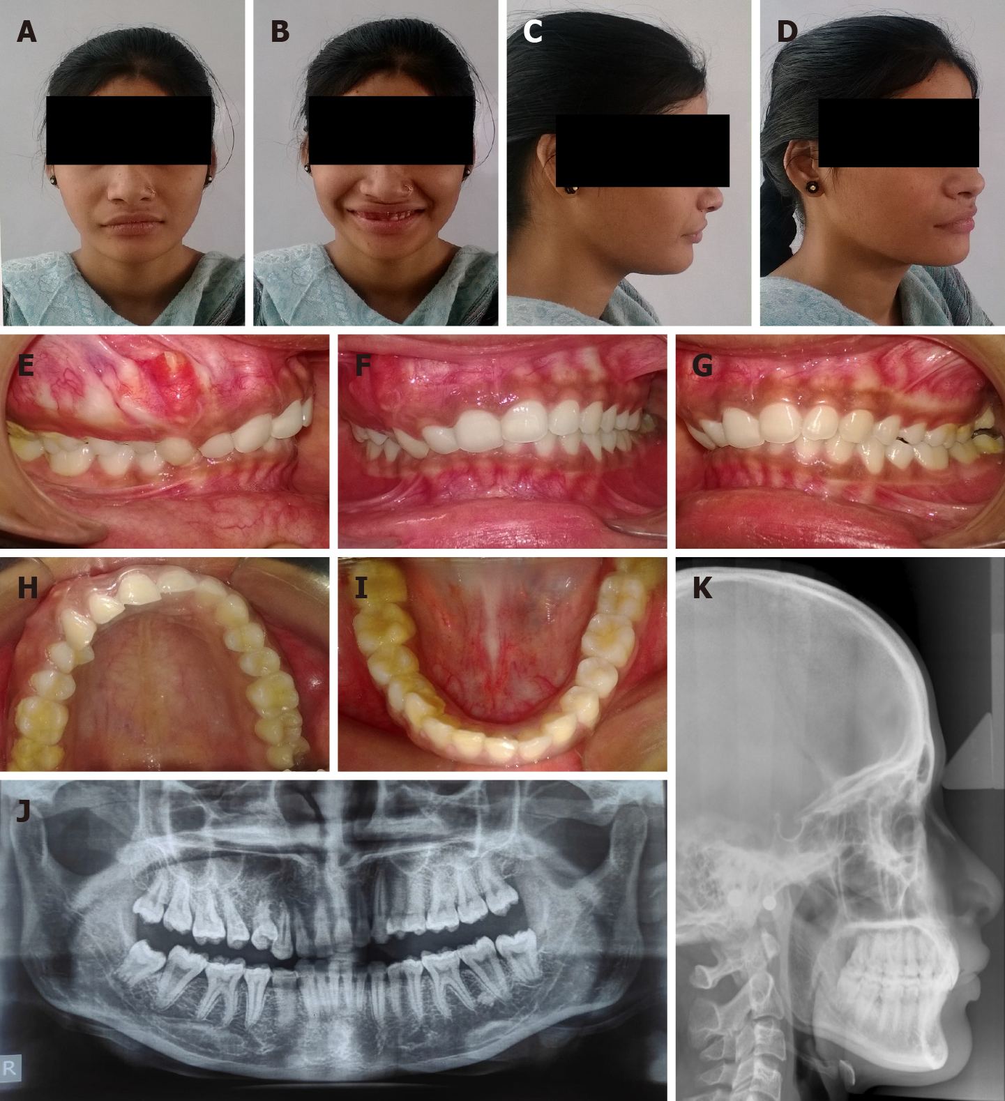
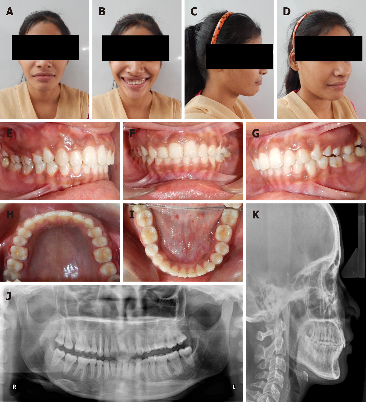
Figure 2 Post-surgical/pre-treatment photographs (October 2013).
A: Extra-oral frontal view; B: Extra-oral frontal with smile view; C: Extra-oral right profile view; D: Extra-oral right three-quarter view; E: Intra-oral right buccal view; F: Intra-oral front view; G: Intra-oral left buccal view; H: Intra-oral maxillary occlusal view; I: Intra-oral mandibular occlusal view; J: Orthopantomogram X-ray; K: Lateral cephalogram.
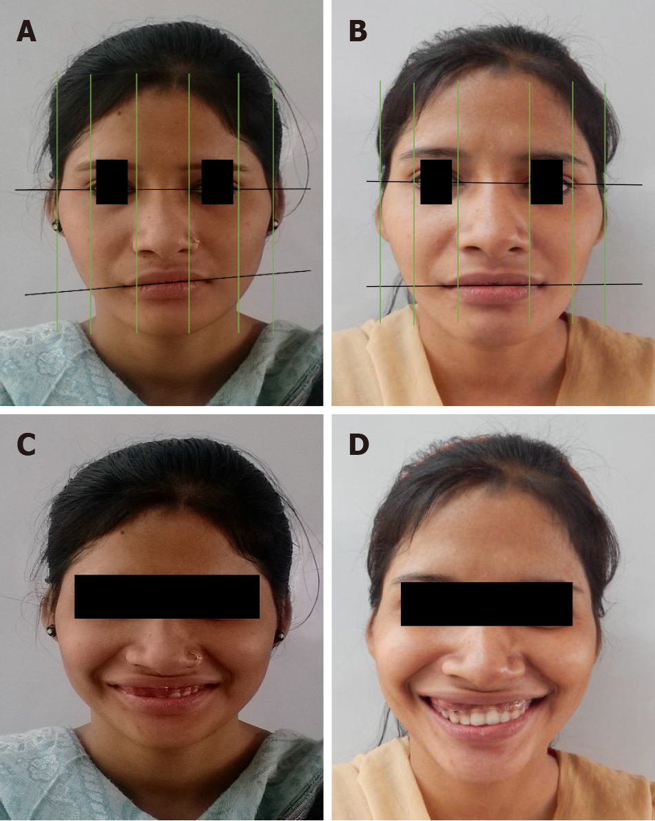
Figure 3 Photographic analysis and pre and post orthodontic treatment comparison.
A and B: Comparison of frontal photographs portray the change in inter-commissural cant resulting from the correction of occlusal cant; C and D: Comparison of smile photographs depict the improvement of smile esthetics resulting from the intrusion of teeth on the affected side.
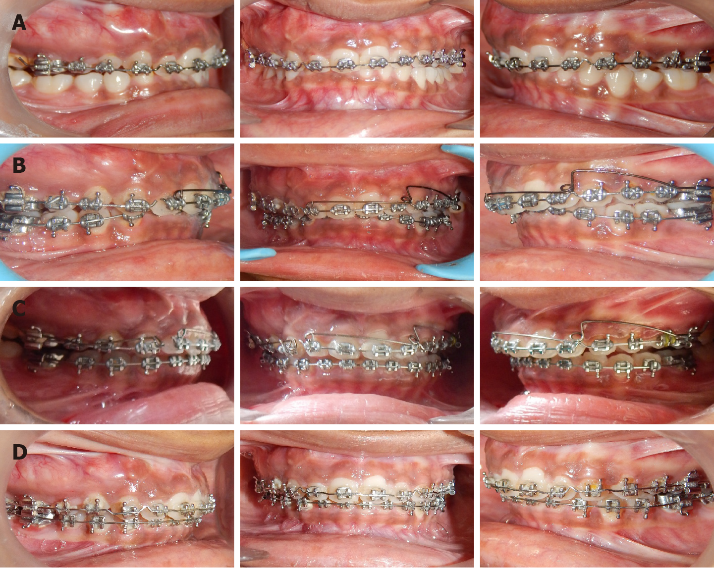
Figure 4 Treatment progress and orthodontic mechanics applied (timeline: Dec 2013-May 2017).
A: Torquing wire with anterior differential palatal root torque; B: Lower arch bonding. Upper arch asymmetric anterior intrusion using unilateral Intrusion arch (.018” AJW); C: Separate canine intrusion arch (.018” AJW) and continued asymmetric anterior intrusion; D: Post-intrusion finishing and detailing.
Figure 5 Post-orthodontic treatment photographs (June 2017).
A: Extra-oral frontal view; B: Extra-oral frontal with smile view; C: Extra-oral right profile view; D: Extra-oral right three-quarter view; E: Intra-oral right buccal view; F: Intra-oral front view; G: Intra-oral left buccal view; H: Intra-oral maxillary occlusal view; I: Intra-oral mandibular occlusal view; J: Orthopantomogram X-ray; K: Lateral cephalogram.
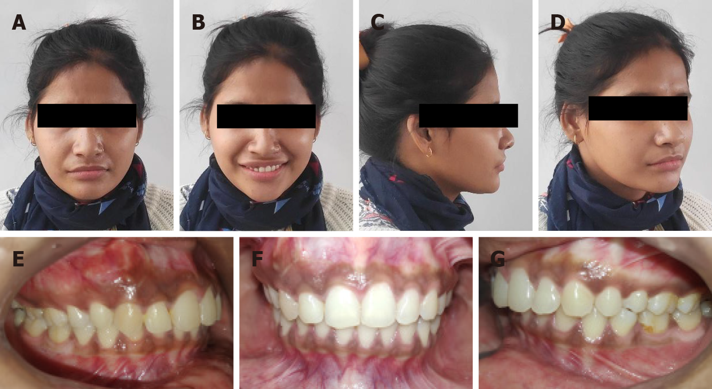
Figure 6 Three years post-retention pics (December 2020).
A: Extra-oral frontal view; B: Extra-oral frontal with smile view; C: Extra-oral right profile view; D: Extra-oral right three-quarter view; E: Intra-oral right buccal view; F: Intra-oral front view; G: Intra-oral left buccal view.
- Citation: Kaur H, Mohanty S, Kochhar GK, Iqbal S, Verma A, Bhasin R, Kochhar AS. Comprehensive management of malocclusion in maxillary fibrous dysplasia: A case report. World J Clin Cases 2021; 9(34): 10671-10680
- URL: https://www.wjgnet.com/2307-8960/full/v9/i34/10671.htm
- DOI: https://dx.doi.org/10.12998/wjcc.v9.i34.10671


