Copyright
©The Author(s) 2021.
World J Clin Cases. Dec 6, 2021; 9(34): 10666-10670
Published online Dec 6, 2021. doi: 10.12998/wjcc.v9.i34.10666
Published online Dec 6, 2021. doi: 10.12998/wjcc.v9.i34.10666
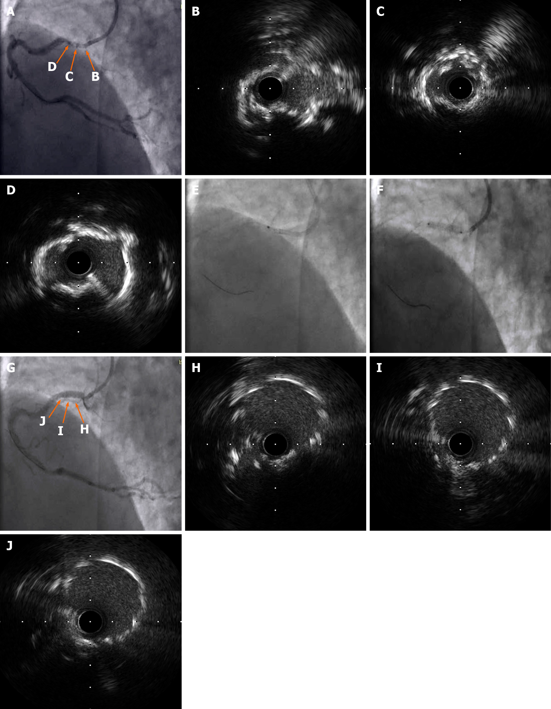
Figure 1 Intravascular ultrasonography findings.
A–D: Intravascular ultrasonography (IVUS) was performed after the laser catheter passed through the lesion and severe calcifications were noted; E and F: The laser catheter was slowly passed through the lesion and the stent was placed at 12 atm; G-J: The final IVUS findings showed no apparent dissection, malapposition, or underexpansion.
- Citation: Hou FJ, Ma XT, Zhou YJ, Guan J. Excimer laser coronary atherectomy for a severe calcified coronary ostium lesion: A case report. World J Clin Cases 2021; 9(34): 10666-10670
- URL: https://www.wjgnet.com/2307-8960/full/v9/i34/10666.htm
- DOI: https://dx.doi.org/10.12998/wjcc.v9.i34.10666


