Copyright
©The Author(s) 2021.
World J Clin Cases. Dec 6, 2021; 9(34): 10645-10651
Published online Dec 6, 2021. doi: 10.12998/wjcc.v9.i34.10645
Published online Dec 6, 2021. doi: 10.12998/wjcc.v9.i34.10645
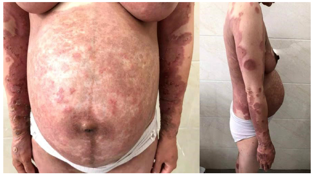
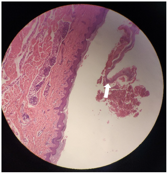
Figure 1 Pemphigoid gestationis on the abdomen and limbs.
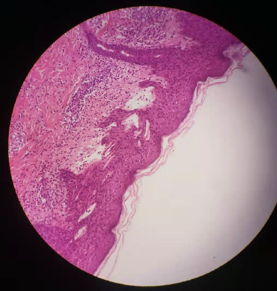
Figure 2 Skin with a sub-epidermal blister with eosinophils and lymphocytes within the cavity and in superficial dermis of pemphigoid gestationis in pregnancy (hematoxylin and eosin 100×).
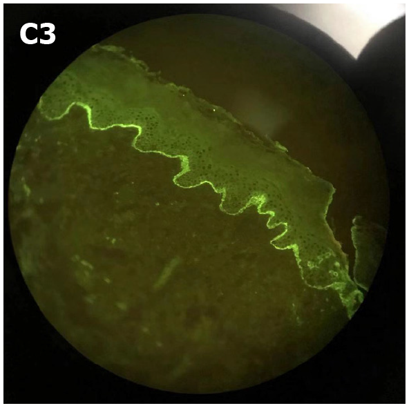
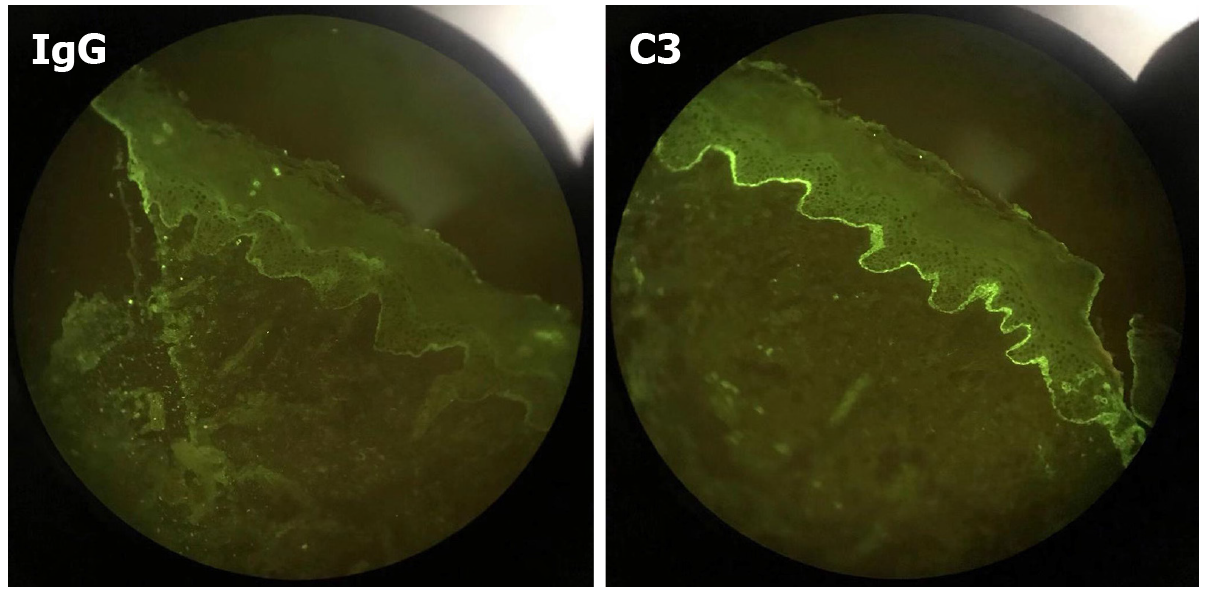
Figure 3 Linear deposition of C3 along the basement membrane zone with the absence of IgG (direct immunofluorescence) of pemphigoid gestationis in pregnancy.
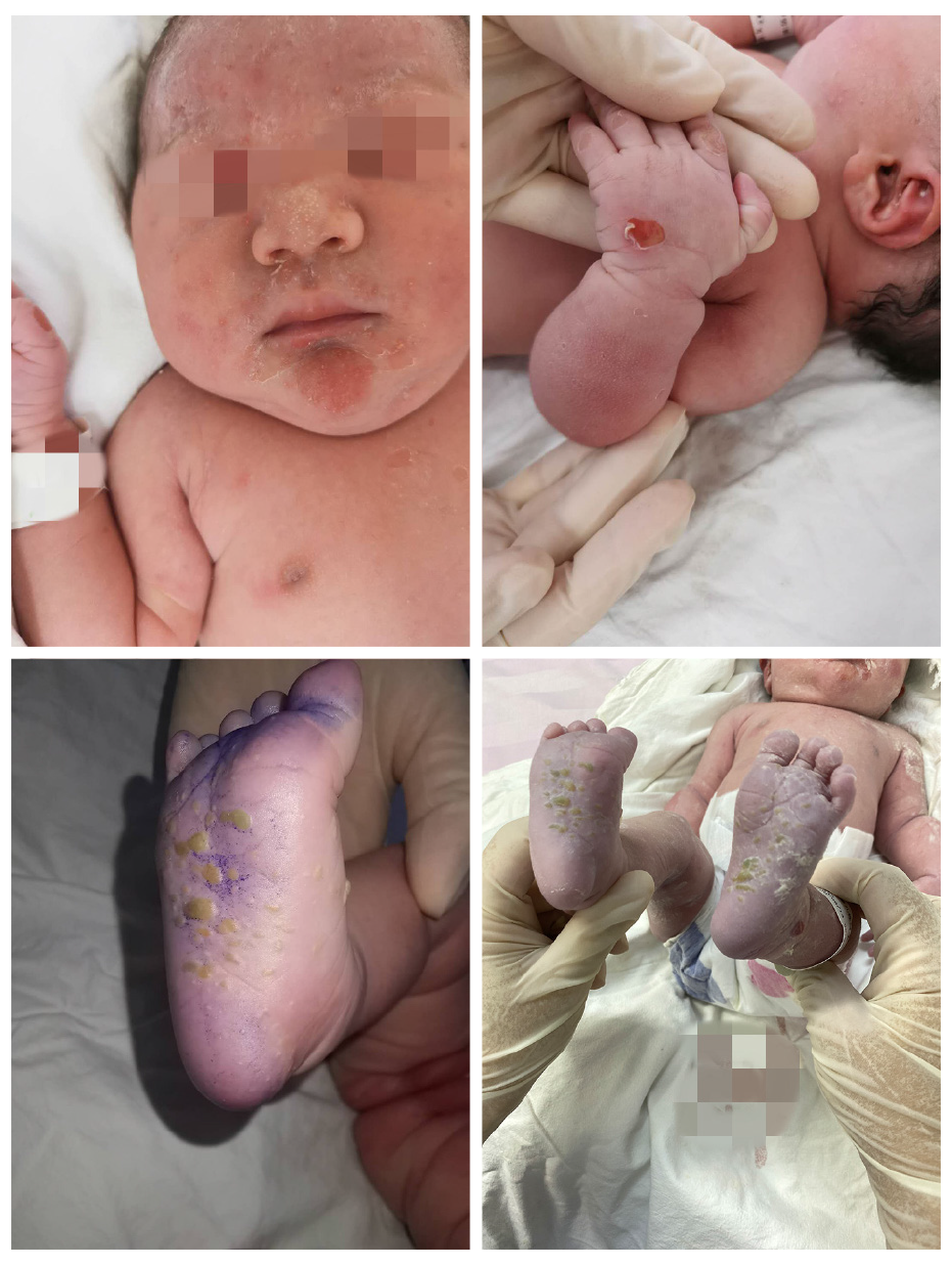
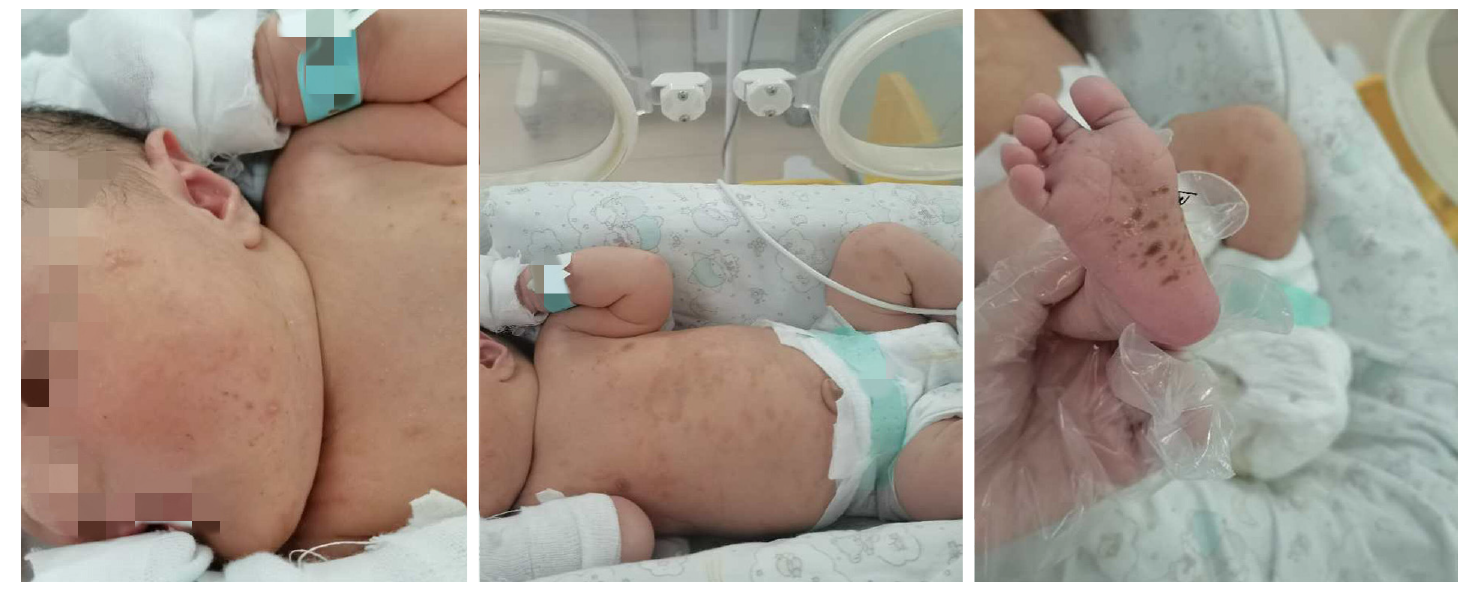
Figure 4 The first day after birth of the infant: Urticaria-like and vesicular skin lesions in neonatal pemphigoid gestationis.
Figure 5 Skin biopsy of the infant showed another layer of epithelium over the normal epithelium with eosinophils and lymphocytes infiltration.
Figure 6 Direct immunofluorescence of the infant showed linear deposits of C3 along the basement membrane zone similar to her mother.
Figure 7 The seventh day after birth of the infant.
- Citation: Jiao HN, Ruan YP, Liu Y, Pan M, Zhong HP. Diagnosis, fetal risk and treatment of pemphigoid gestationis in pregnancy: A case report. World J Clin Cases 2021; 9(34): 10645-10651
- URL: https://www.wjgnet.com/2307-8960/full/v9/i34/10645.htm
- DOI: https://dx.doi.org/10.12998/wjcc.v9.i34.10645


