Copyright
©The Author(s) 2021.
World J Clin Cases. Nov 6, 2021; 9(31): 9686-9690
Published online Nov 6, 2021. doi: 10.12998/wjcc.v9.i31.9686
Published online Nov 6, 2021. doi: 10.12998/wjcc.v9.i31.9686
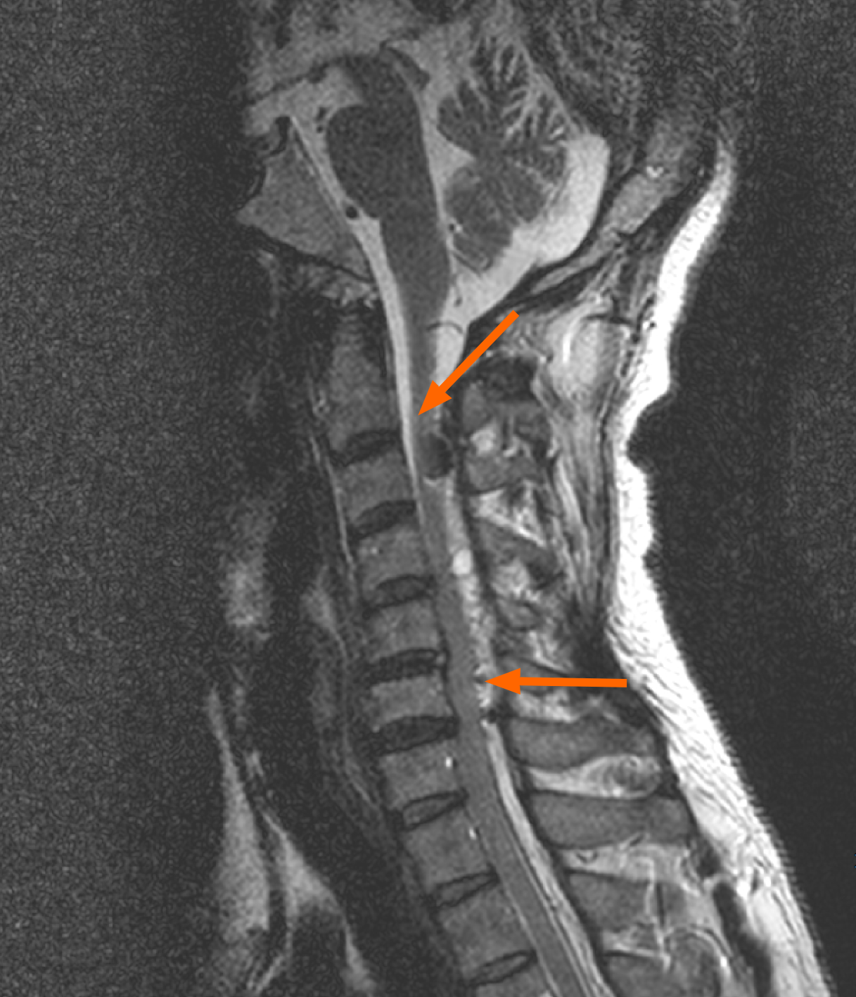
Figure 1 T2-weighted sagittal cervical magnetic resonance image demonstrating an epidural hemorrhage with gas bubbles from C2 to the upper thoracic level, resulting in central spinal canal stenosis and cord compression at the C3-T1 level (orange arrows).
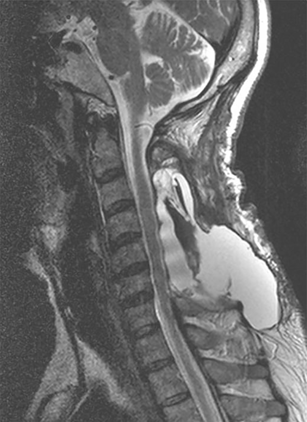
Figure 2 T2-weighted magnetic resonance image demonstrating fluid collection (5.
6 cm × 6.6 cm × 11.2 cm) at the laminectomy site and in the posterior soft tissue at the C3-T1 level.
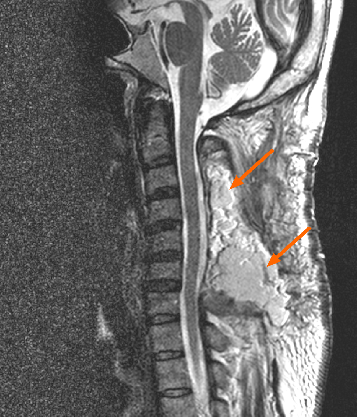
Figure 3 T2-weighted magnetic resonance image demonstrating a repaired pseudomeningocele and an abdominal vascularized fat graft transplantation (orange arrows).
- Citation: Kim KW, Cho JH. Iatrogenic giant pseudomeningocele of the cervical spine: A case report. World J Clin Cases 2021; 9(31): 9686-9690
- URL: https://www.wjgnet.com/2307-8960/full/v9/i31/9686.htm
- DOI: https://dx.doi.org/10.12998/wjcc.v9.i31.9686


