Copyright
©The Author(s) 2021.
World J Clin Cases. Nov 6, 2021; 9(31): 9645-9651
Published online Nov 6, 2021. doi: 10.12998/wjcc.v9.i31.9645
Published online Nov 6, 2021. doi: 10.12998/wjcc.v9.i31.9645
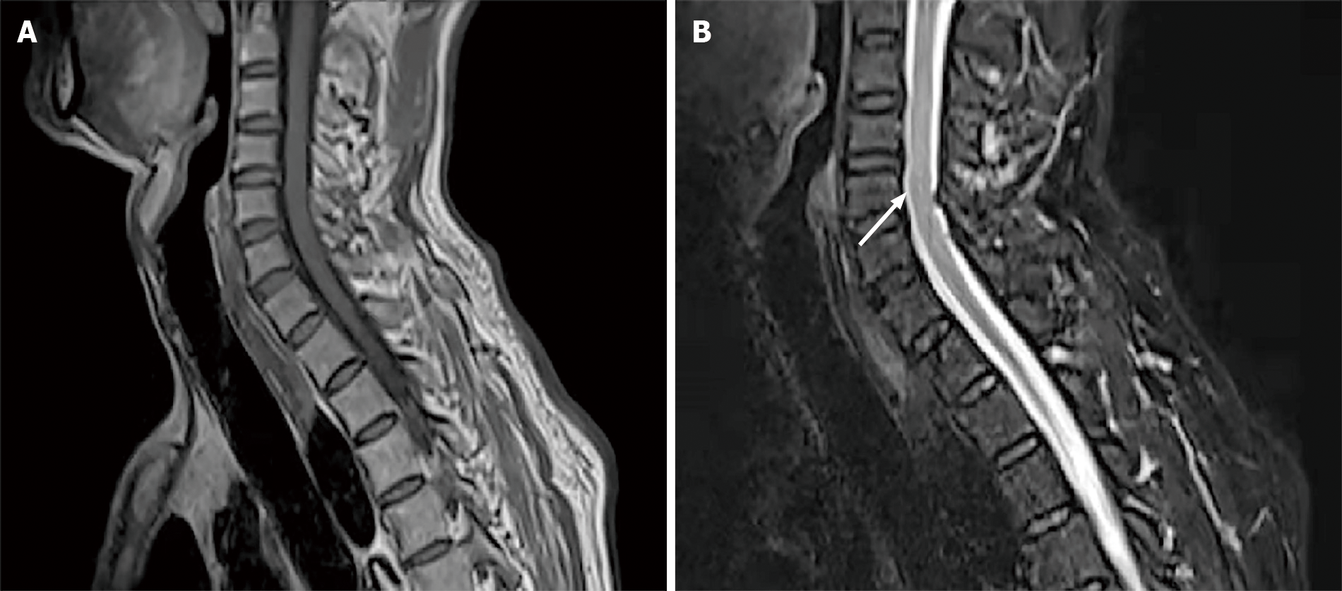
Figure 1 Thoracic spinal magnetic resonance imaging (MRI) on June 5, 2020.
MRI showed multiple lesions along the cervicothoracic junction (arrow). A: T1-weighted sequence; B: T2-weighted sequence.
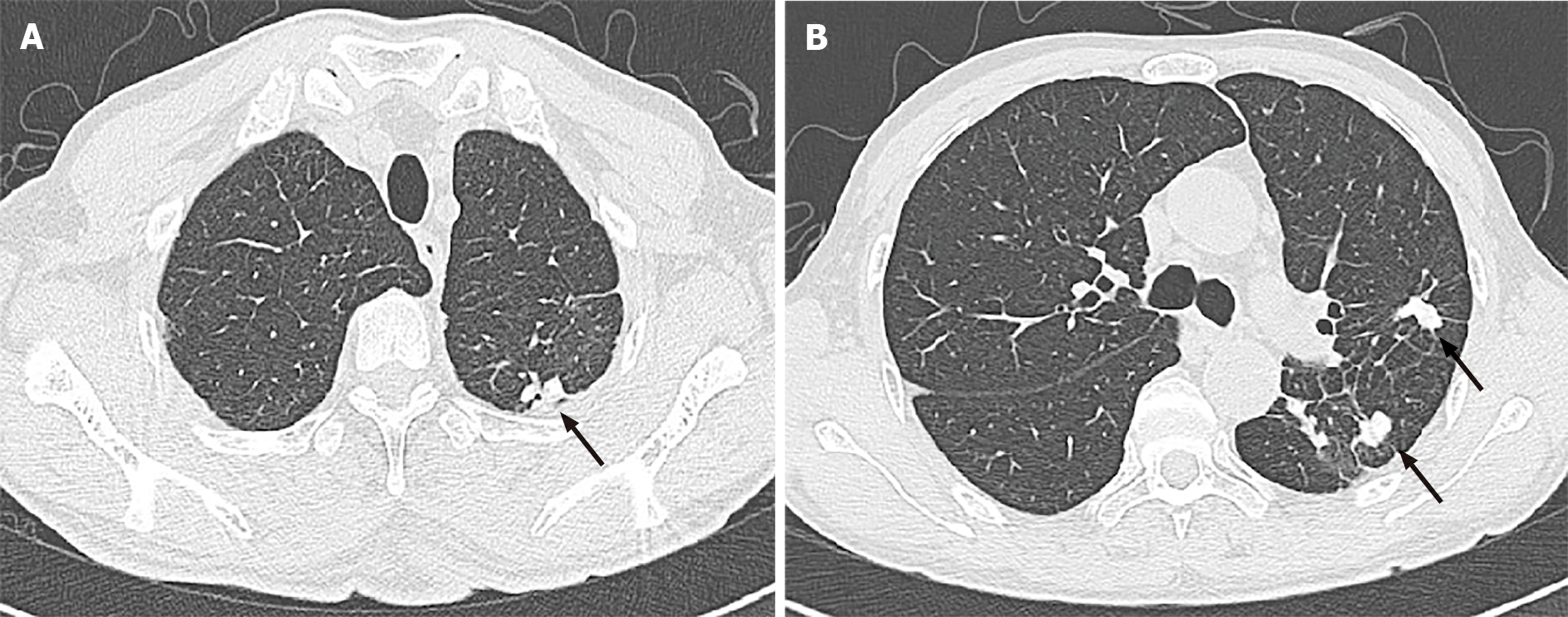
Figure 2 Chest computed tomography conducted on June 5, 2020.
The images showed patchy TB lesions in the left lung (arrows). A: Apical posterior segment of the upper lobe; B: Dorsal segment of the lower lobe.
- Citation: Gu LY, Tian J, Yan YP. Concurrent tuberculous transverse myelitis and asymptomatic neurosyphilis: A case report. World J Clin Cases 2021; 9(31): 9645-9651
- URL: https://www.wjgnet.com/2307-8960/full/v9/i31/9645.htm
- DOI: https://dx.doi.org/10.12998/wjcc.v9.i31.9645


