Copyright
©The Author(s) 2021.
World J Clin Cases. Nov 6, 2021; 9(31): 9629-9634
Published online Nov 6, 2021. doi: 10.12998/wjcc.v9.i31.9629
Published online Nov 6, 2021. doi: 10.12998/wjcc.v9.i31.9629
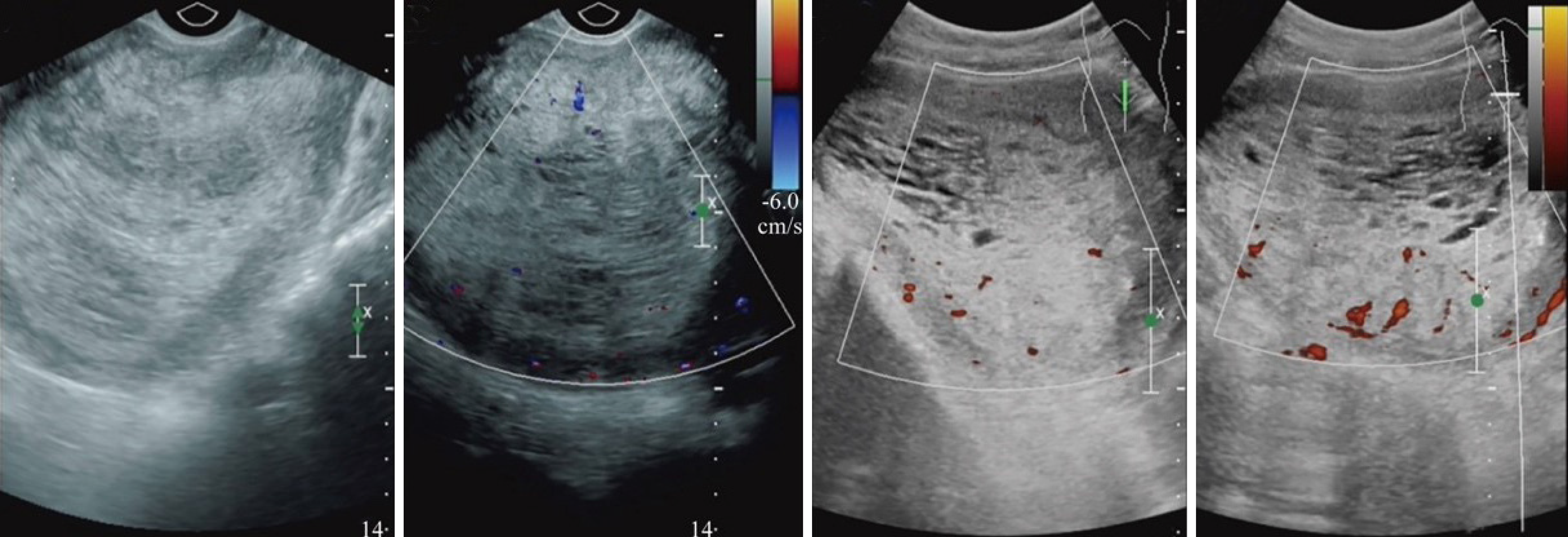
Figure 1 The normal uterine cavity line was not clear along the direction of the uterine cavity to explore the cluster that was approximately 10.
1 cm × 7.5 cm × 8.7 cm in size. There were a number of different sizes in the dark liquid area, as part of the honeycomb-like appearance, and the light and uterine wall had a boundary. The cervical canal extended in the downward direction.
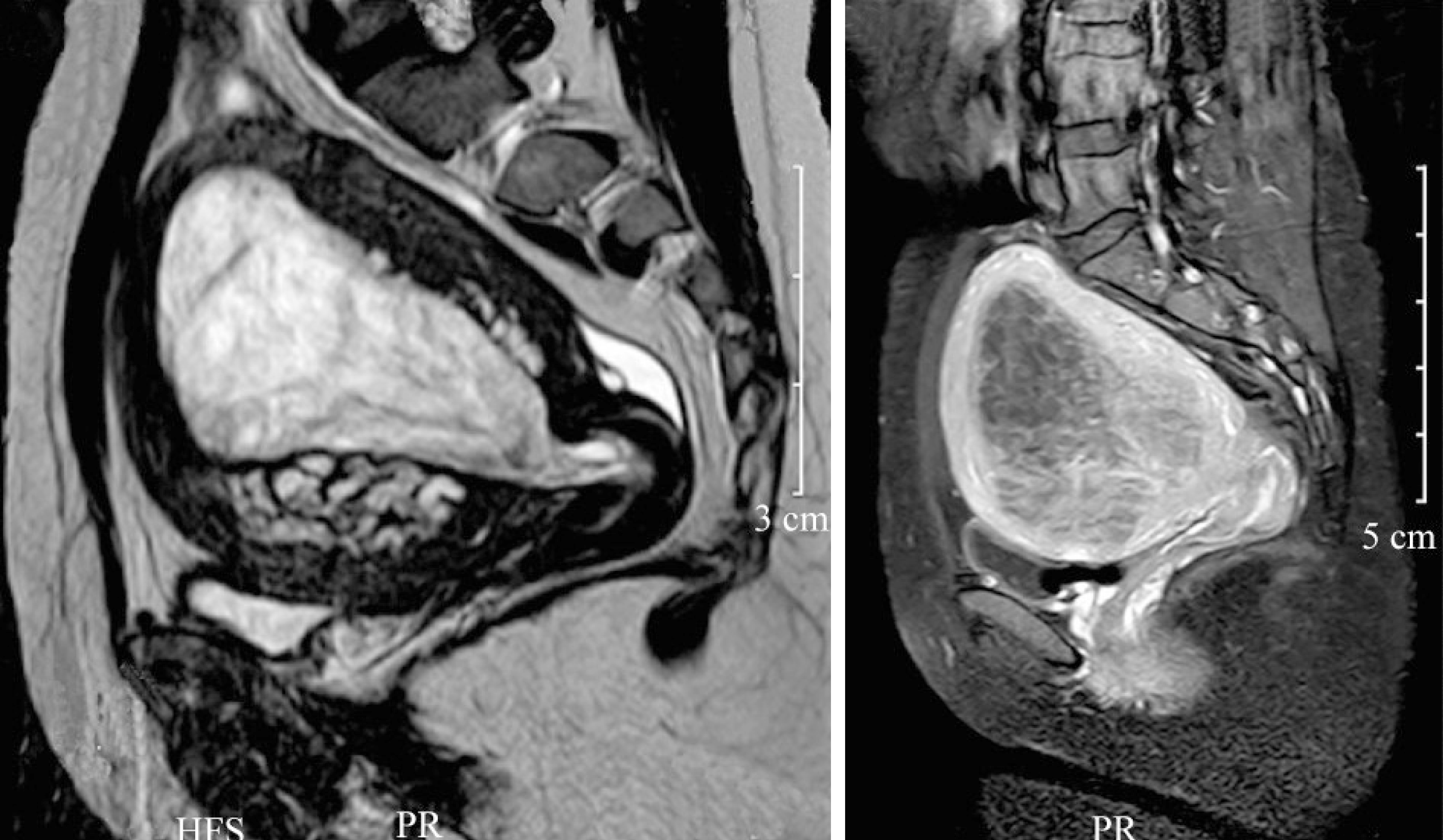
Figure 2 Focus and intensity of the abnormal signal in the intrauterine cavity, which was approximately 81 mm × 82 mm × 91 mm in size.
The invasion and nature of the myometrium were determined to be an invasive hydatidiform mole.
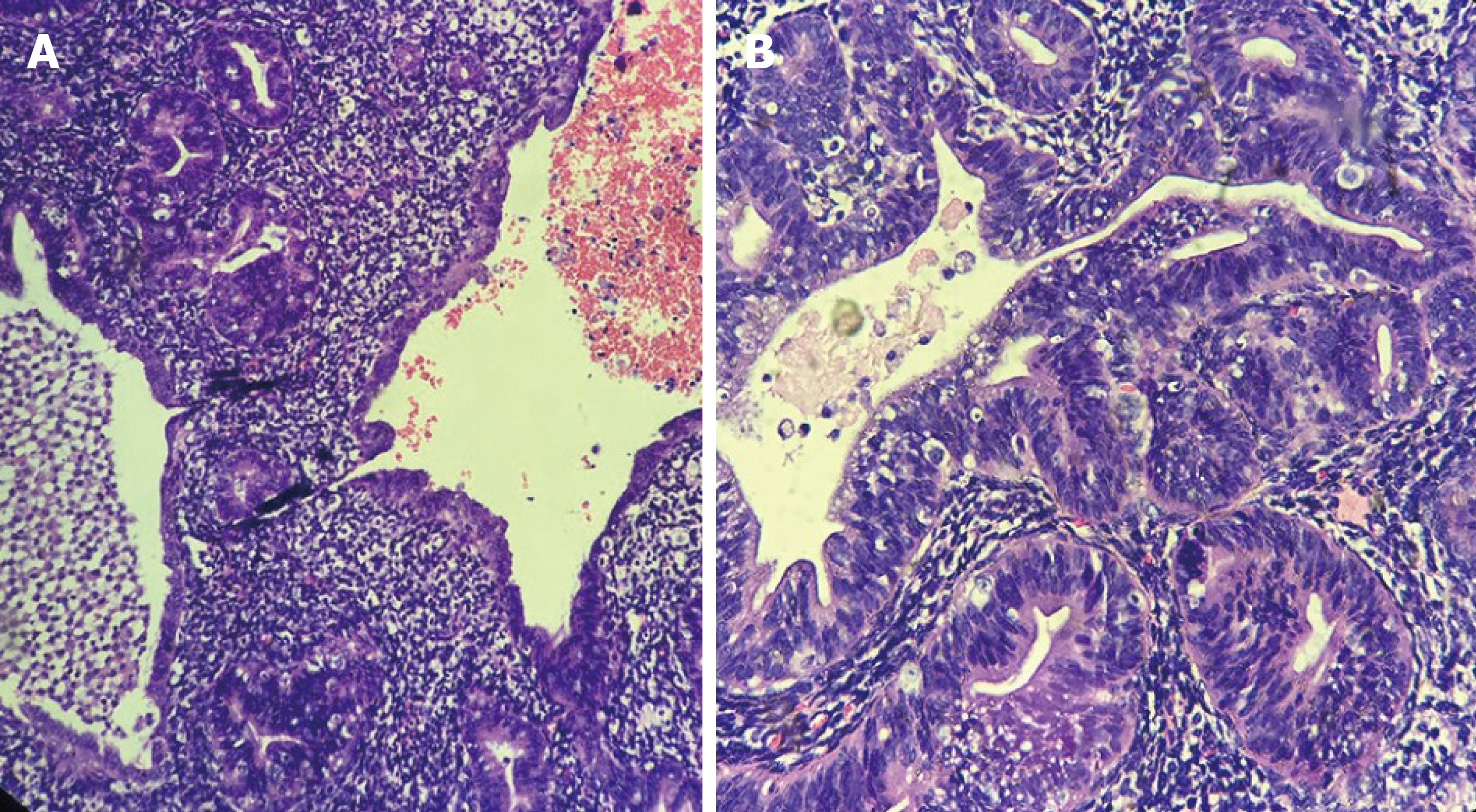
Figure 3 Endometrial hyperplasia, foci with atypical hyperplasia.
A: Hematoxylin and eosin (HE) staining, × 100 magnification; B: HE staining, × 200 magnification.
- Citation: Wu X, Luo J, Wu F, Li N, Tang AQ, Li A, Tang XL, Chen M. Atypical endometrial hyperplasia in a 35-year-old woman: A case report and literature review. World J Clin Cases 2021; 9(31): 9629-9634
- URL: https://www.wjgnet.com/2307-8960/full/v9/i31/9629.htm
- DOI: https://dx.doi.org/10.12998/wjcc.v9.i31.9629


