Copyright
©The Author(s) 2021.
World J Clin Cases. Nov 6, 2021; 9(31): 9617-9622
Published online Nov 6, 2021. doi: 10.12998/wjcc.v9.i31.9617
Published online Nov 6, 2021. doi: 10.12998/wjcc.v9.i31.9617
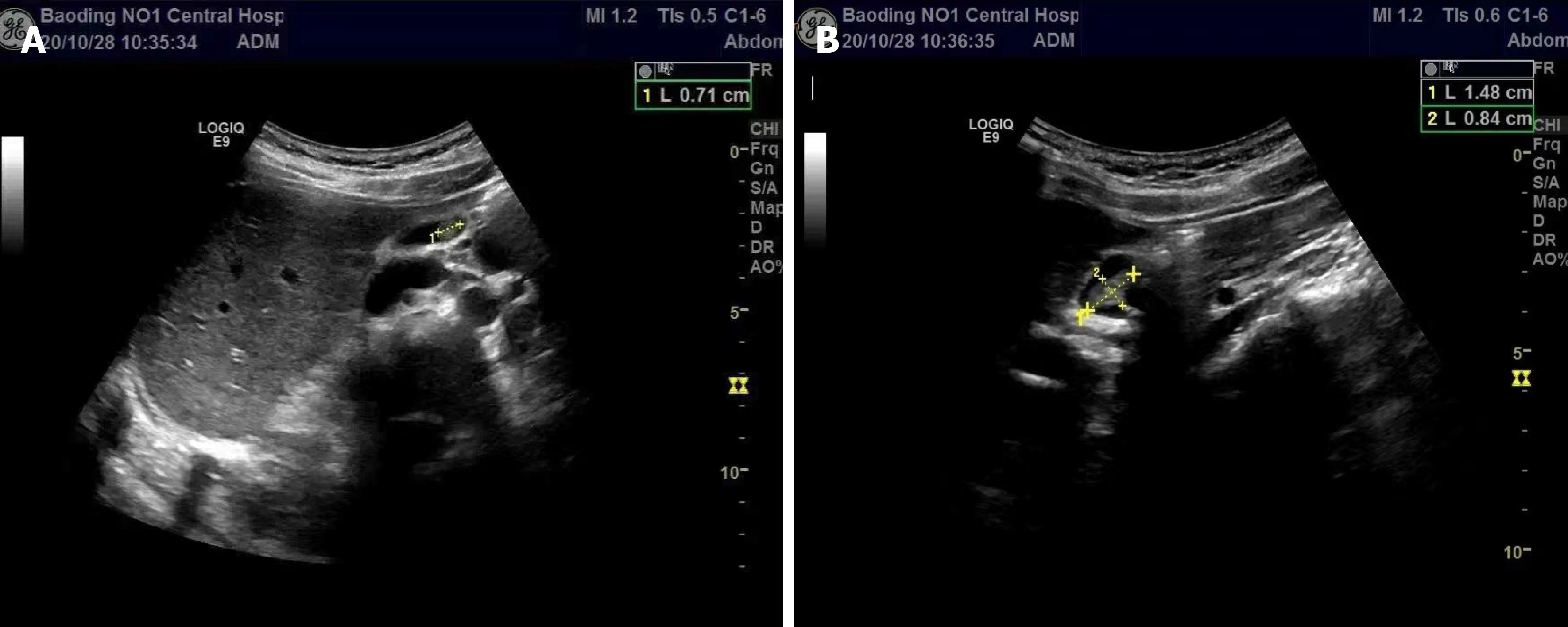
Figure 1 The polyp in the gallbladder shows a medium-strong echo intensity.
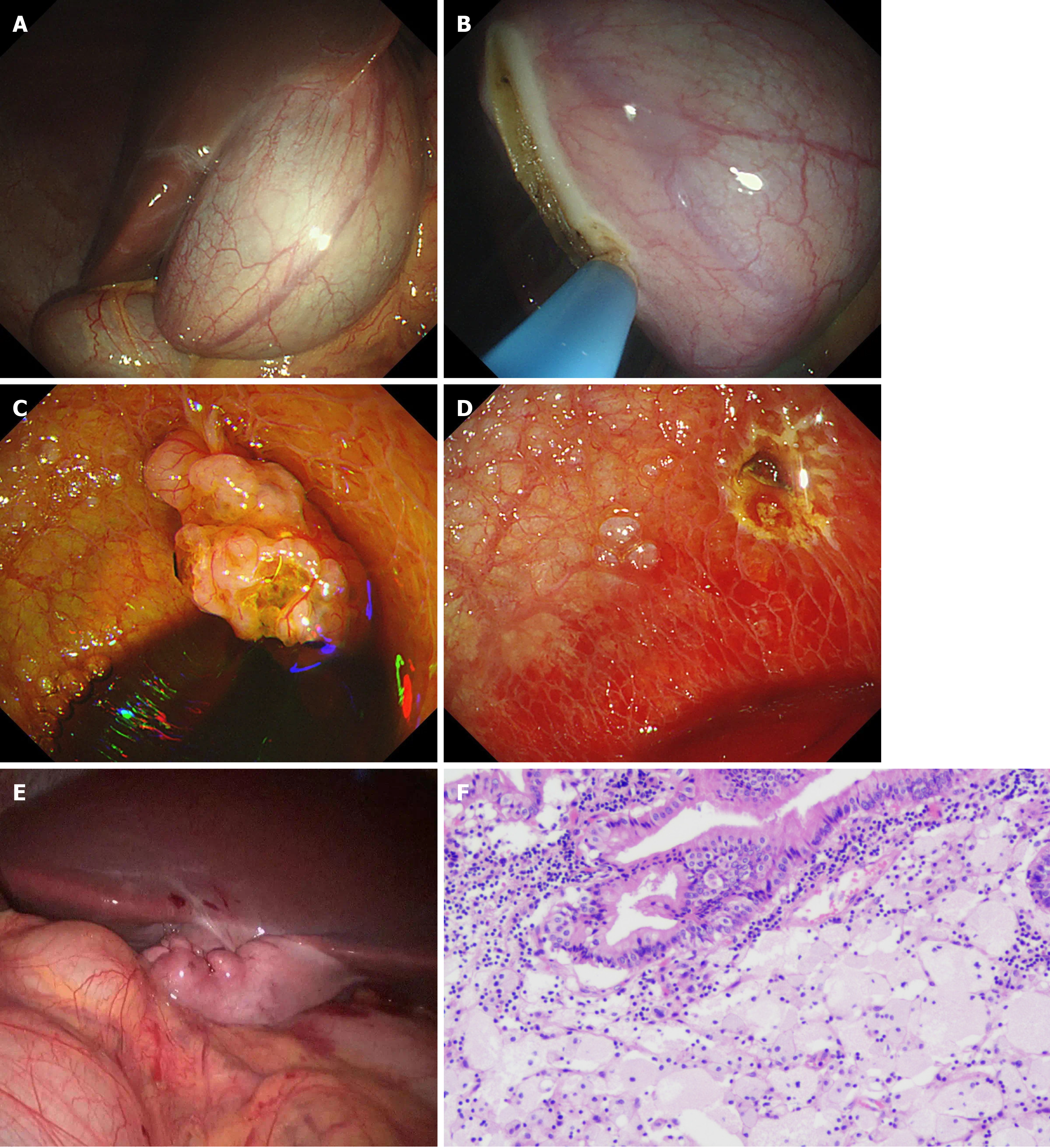
Figure 2 Laparoscopic-assisted transumbilical gastroscopy for gallbladder-preserving polypectomy.
A and B: The gastroscope entered the abdominal cavity through the umbilical trocar. The gallbladder was located with the assistance of laparoscopy, and an approximately 2-cm incision in the gallbladder wall was made longitudinally at the bottom of the gallbladder; C and D: Endoscopy entering the gallbladder showed a pedicled polyp with the appearance of a grape cluster. The lesion was resected by high-frequency electroresection; active bleeding could be seen at the base, and haemostasis was achieved by electrocoagulation with haemostatic forceps. The specimen was removed and fixed for pathological examination; E: The gastroscope was withdrawn from the abdominal cavity. Laparoscopic suture of the gallbladder incision was performed, followed by full flushing of the abdominal pelvic cavity and suture of the abdominal incision; F: The specimen was found to be a cholesterol polyp (Original magnification 200 ×).
- Citation: Zheng Q, Zhang G, Yu XH, Zhao ZF, Lu L, Han J, Zhang JZ, Zhang JK, Xiong Y. Perfect pair, scopes unite — laparoscopic-assisted transumbilical gastroscopy for gallbladder-preserving polypectomy: A case report. World J Clin Cases 2021; 9(31): 9617-9622
- URL: https://www.wjgnet.com/2307-8960/full/v9/i31/9617.htm
- DOI: https://dx.doi.org/10.12998/wjcc.v9.i31.9617


