Copyright
©The Author(s) 2021.
World J Clin Cases. Nov 6, 2021; 9(31): 9577-9583
Published online Nov 6, 2021. doi: 10.12998/wjcc.v9.i31.9577
Published online Nov 6, 2021. doi: 10.12998/wjcc.v9.i31.9577
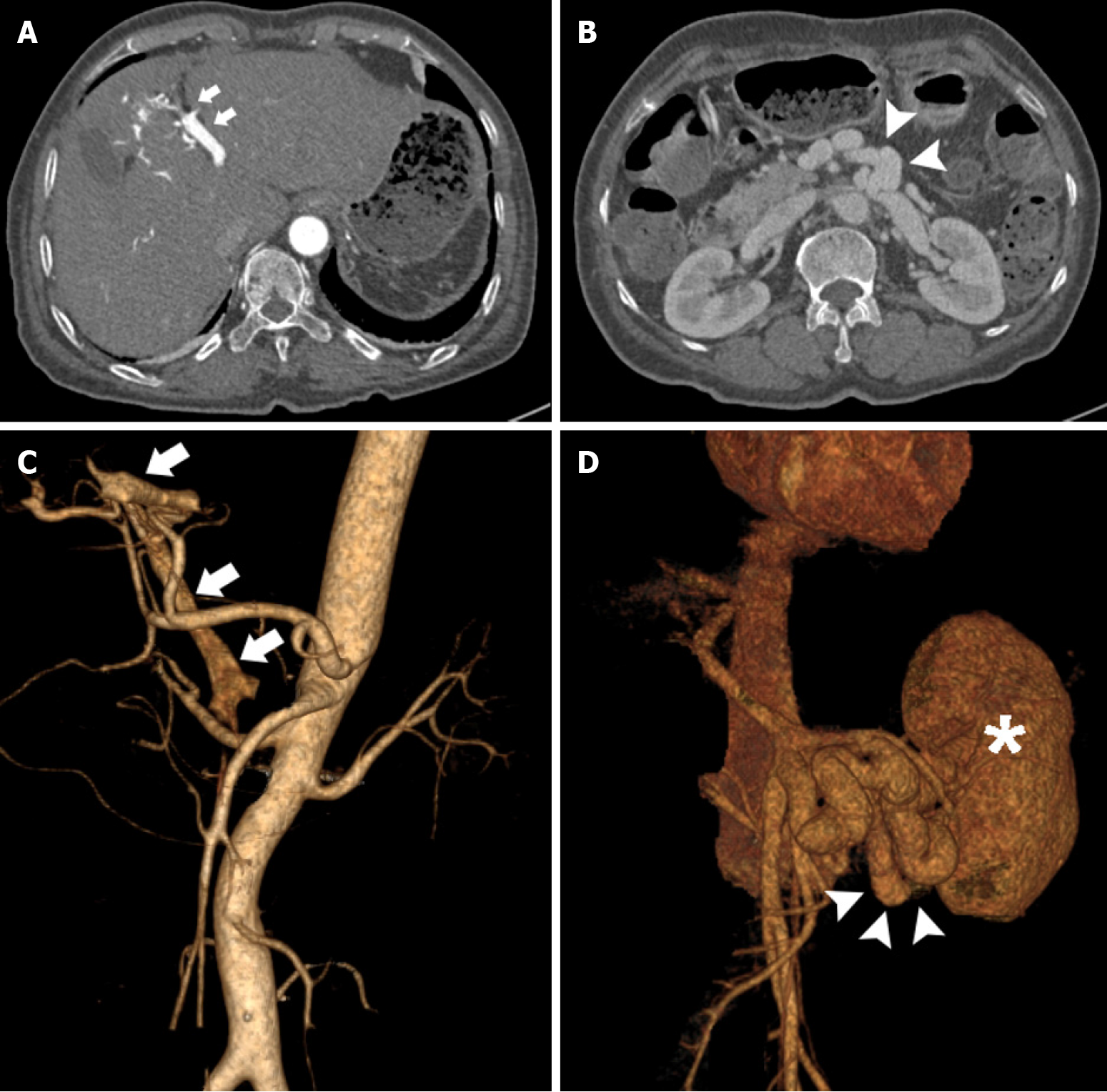
Figure 1 Contrast-enhanced abdominal computed tomography.
A: Computed tomography (CT) revealing early visualization of the portal vein (arrow) in the arterial phase; B: CT showing a large shunt (arrow) between the superior mesenteric vein and inferior vena cava; C: Three-dimensional vascular reconstruction demonstrating an intrahepatic arterioportal fistula (arrow); D: Three-dimensional vascular reconstruction revealing a large spontaneous portosystemic shunt (arrow) beside the left kidney (asterisk).
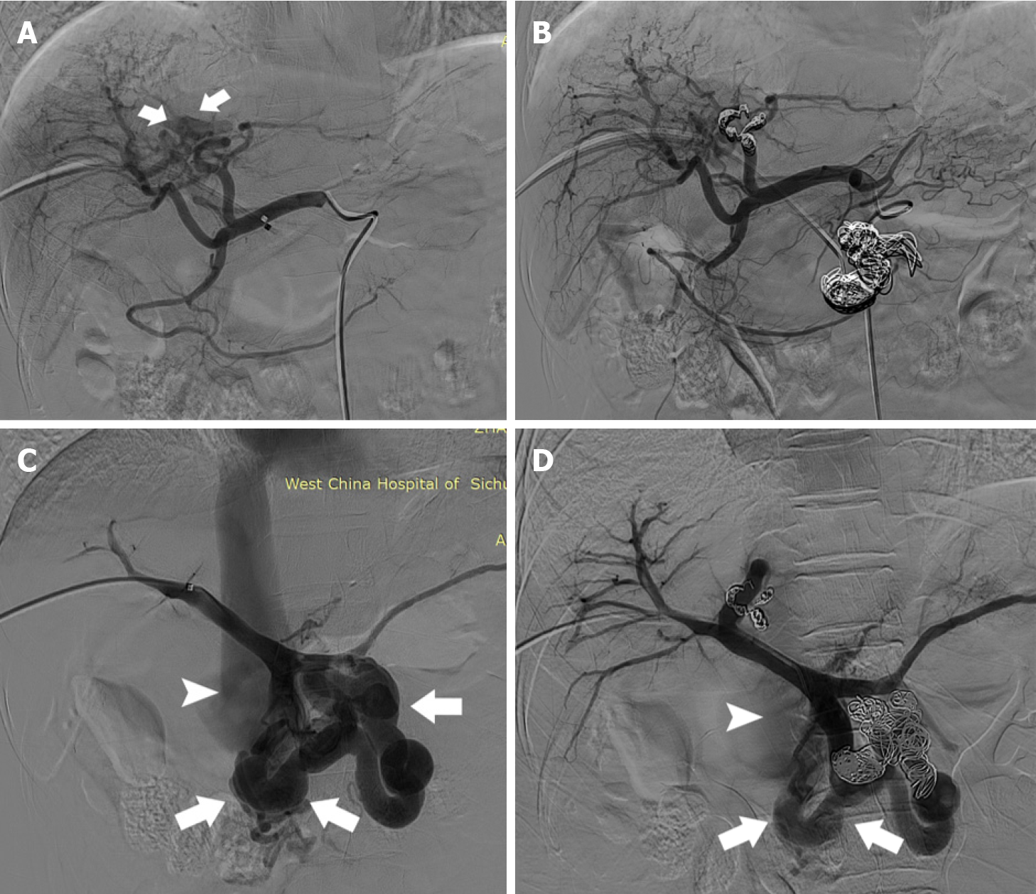
Figure 2 Selective angiography of the common hepatic artery and portal trunk.
A: Selective digital subtraction angiography of the common hepatic artery revealing rapid filling through the fistula (arrow) into the left branch of the portal vein; B: The fistula was abrogated after embolization with stainless metal coils; C: Selective angiography of the portal trunk showing the large shunt between the superior mesenteric vein and inferior vena cava (arrow); D: Selective angiography of the portal trunk demonstrating that the opacification of the shunt had markedly decreased (arrow) after embolization.
- Citation: Liu GF, Wang XZ, Luo XF. Simultaneous embolization of a spontaneous porto-systemic shunt and intrahepatic arterioportal fistula: A case report. World J Clin Cases 2021; 9(31): 9577-9583
- URL: https://www.wjgnet.com/2307-8960/full/v9/i31/9577.htm
- DOI: https://dx.doi.org/10.12998/wjcc.v9.i31.9577


