Copyright
©The Author(s) 2021.
World J Clin Cases. Nov 6, 2021; 9(31): 9564-9570
Published online Nov 6, 2021. doi: 10.12998/wjcc.v9.i31.9564
Published online Nov 6, 2021. doi: 10.12998/wjcc.v9.i31.9564
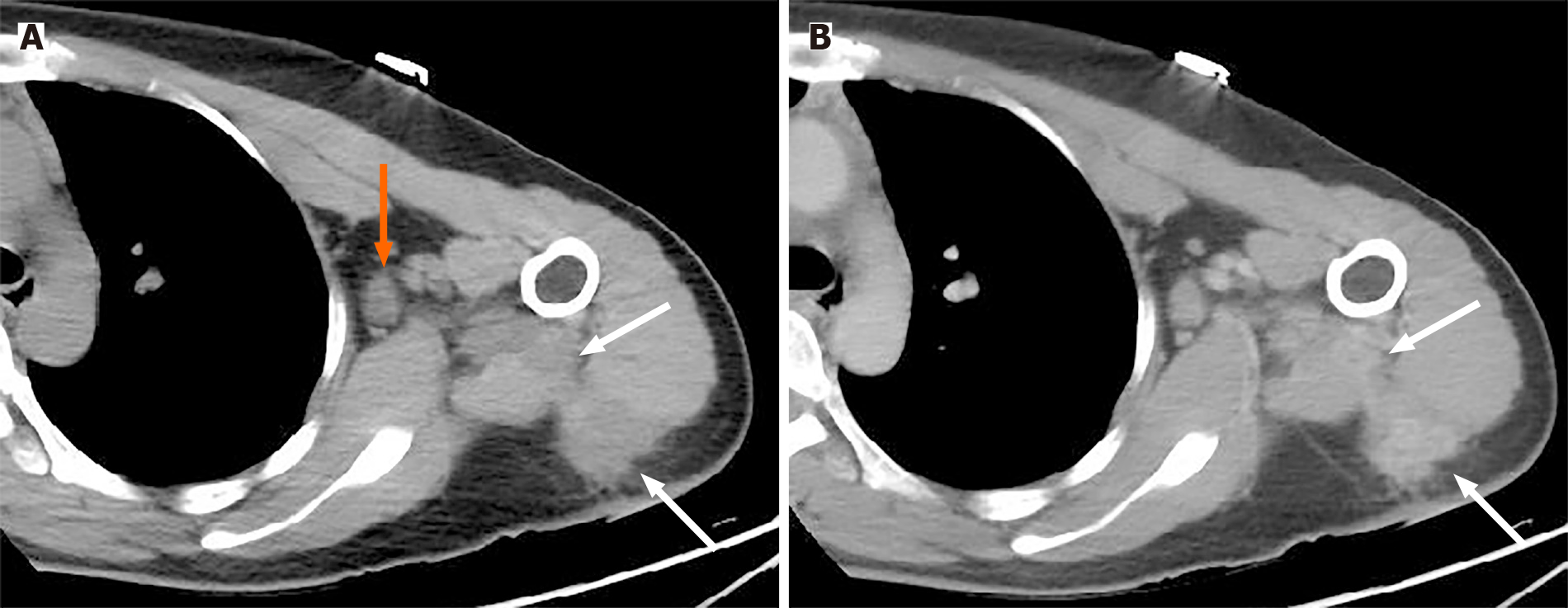
Figure 1 A 36-year-old woman was admitted to our hospital with swelling, skin redness and pain in the upper limb that persisted for 6 mo without a prior history of trauma.
A: Plain computed tomography (CT) imaging shows an irregular hypodense mass in the superficial deltoid muscle extending to the intermuscular space (white arrow) with axillary lymphadenopathy (orange arrow); B: Contrast CT (venous phase) reveals a slightly persistent inhomogeneous enhancement of the mass with a blurred margin (white arrow).
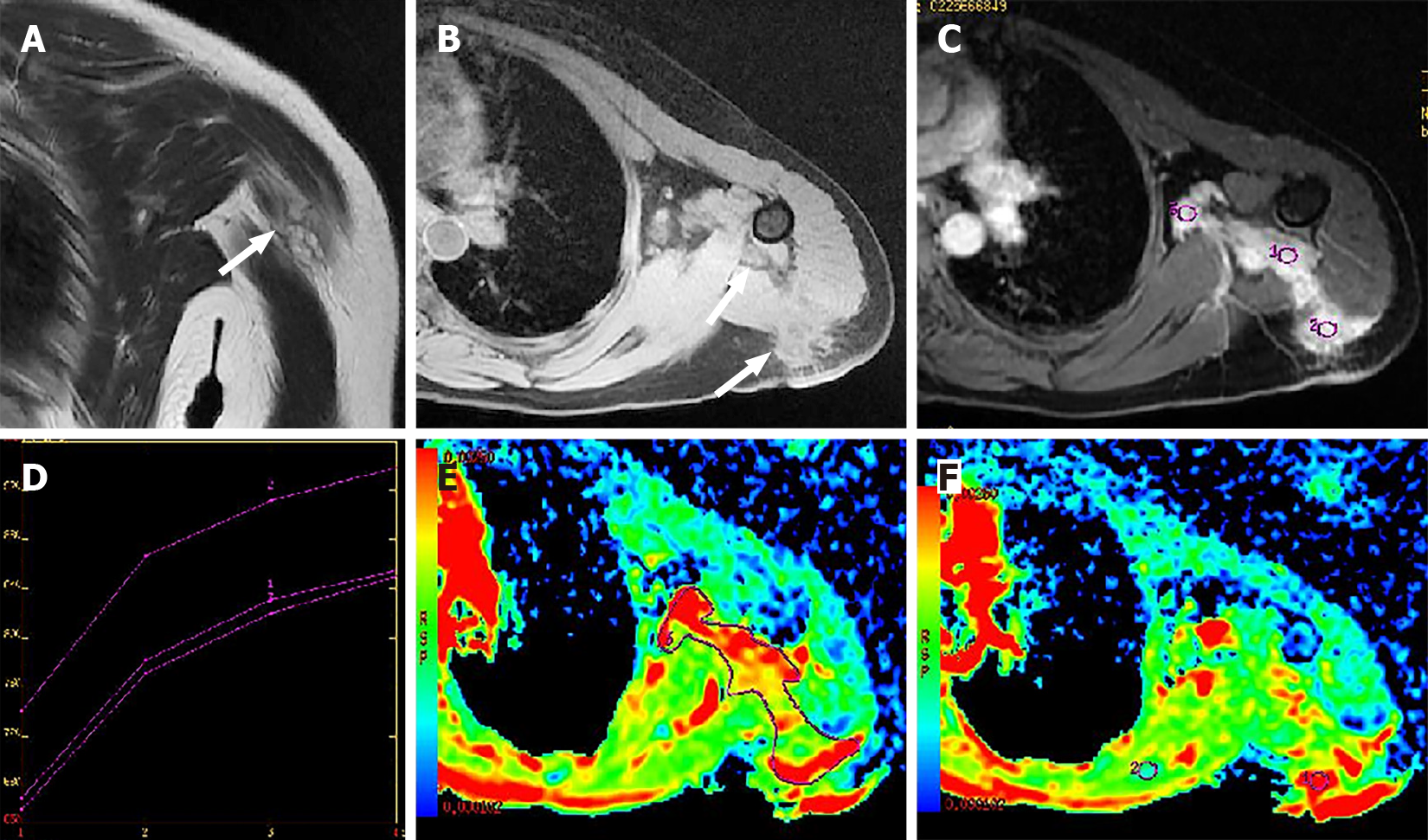
Figure 2 On morphological magnetic resonance images.
Iso- to hyper-intensity of the mass (white arrow) on the coronal T2-weighted without fat saturation image (A) and hypo- to iso-intensity (white arrow) on the axial T1 FSPGR (pre-contrast enhancement) (B) are shown compared to signal intensity. Axial T1 3D FSPGR LAVA sequence shows multiple nodular enhancements with irregular shapes and blurred margins (ROI1, 2, 3) (C). TIC of the mass presents as a slow-increased trend (D). The lesion on DWI was hyperintense with a higher mean ADC value of 2.19 × 10⁻³ mm2/s (E, ROI 3) than surrounding normal soft tissues (1.03 × 10⁻³ mm2/s) (F, ROI2).
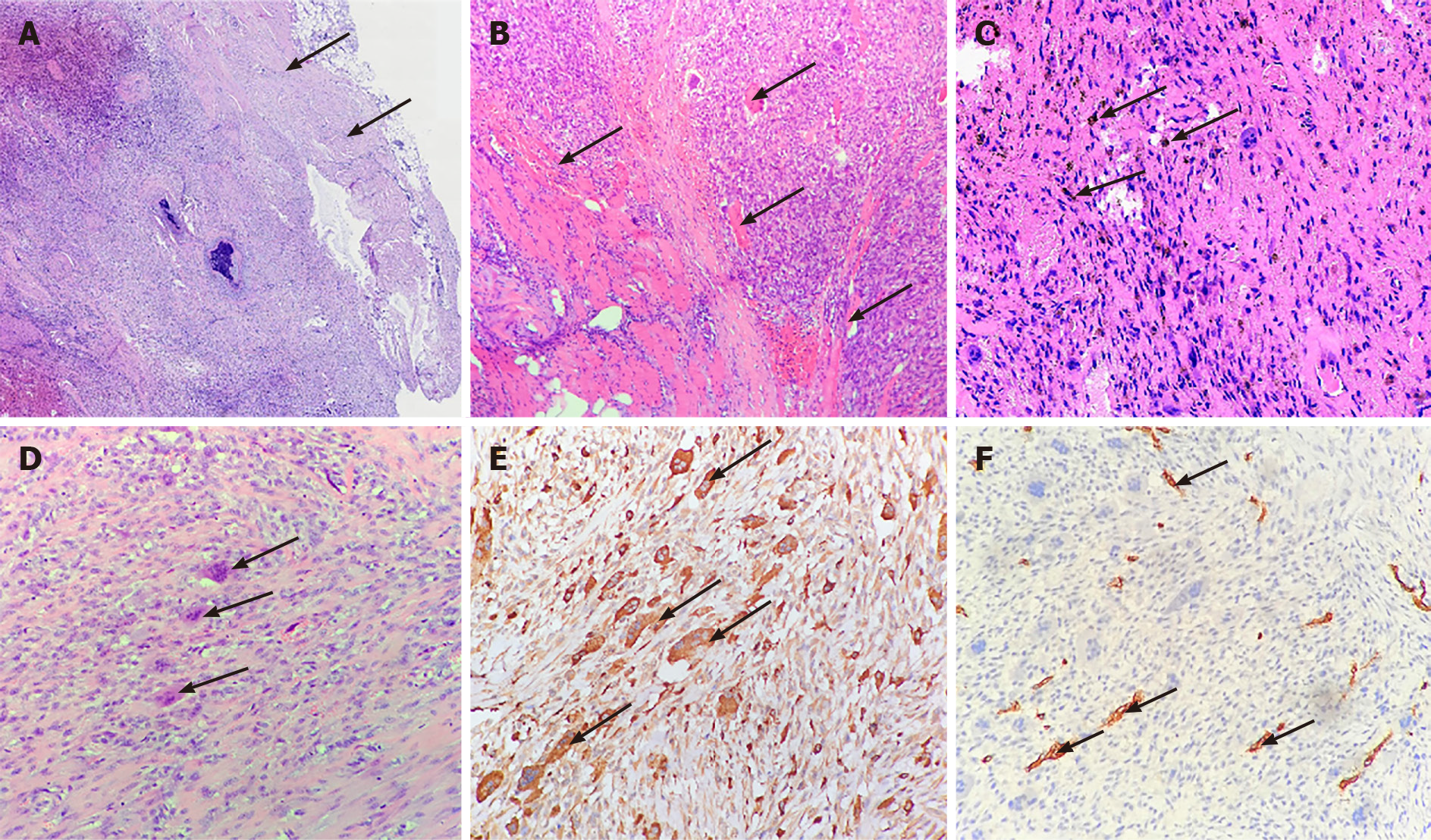
Figure 3 Pathological features of the tumor.
A: The relatively clear boundary (arrows) of the tumor; B: The tumor had invaded neighboring muscles (arrows) on one side; C: Hemosiderin-containing cells (arrows) indicated the tumor with interstitial hemorrhage; D: The tumor had many multinucleated giant cells (arrows) surrounded by spindle cells; E: Multinuclear cells showed strongly positive CD68 (arrows); F: The tumor cells showed negative CD34 but were reactive to vascular endothelial cells (arrows) in the stroma.
- Citation: Kang JY, Zhang K, Liu AL, Wang HL, Zhang LN, Liu WV. Characteristics of primary giant cell tumor in soft tissue on magnetic resonance imaging: A case report. World J Clin Cases 2021; 9(31): 9564-9570
- URL: https://www.wjgnet.com/2307-8960/full/v9/i31/9564.htm
- DOI: https://dx.doi.org/10.12998/wjcc.v9.i31.9564


