Copyright
©The Author(s) 2021.
World J Clin Cases. Nov 6, 2021; 9(31): 9417-9430
Published online Nov 6, 2021. doi: 10.12998/wjcc.v9.i31.9417
Published online Nov 6, 2021. doi: 10.12998/wjcc.v9.i31.9417
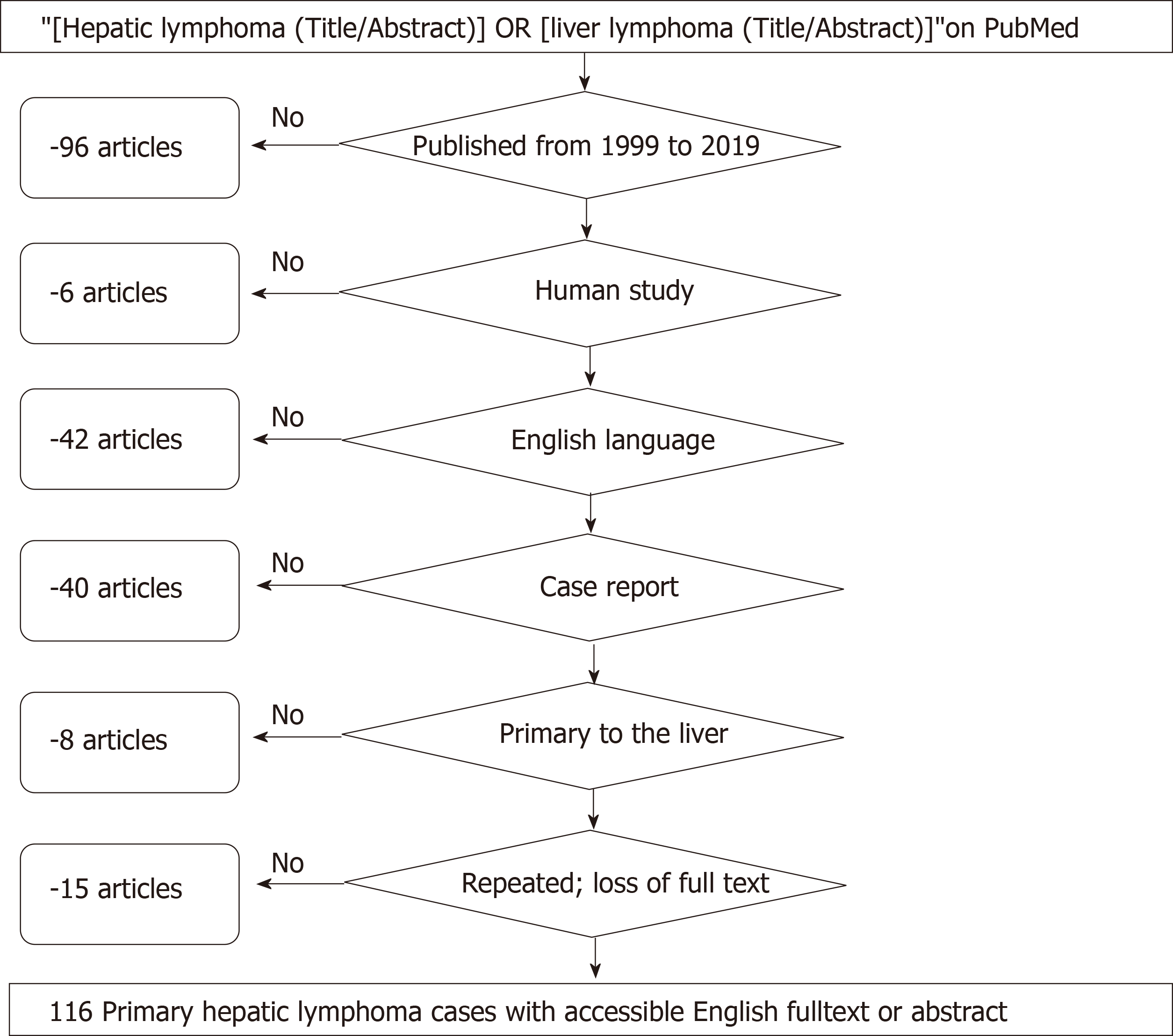
Figure 1
Literature review flow-charts.
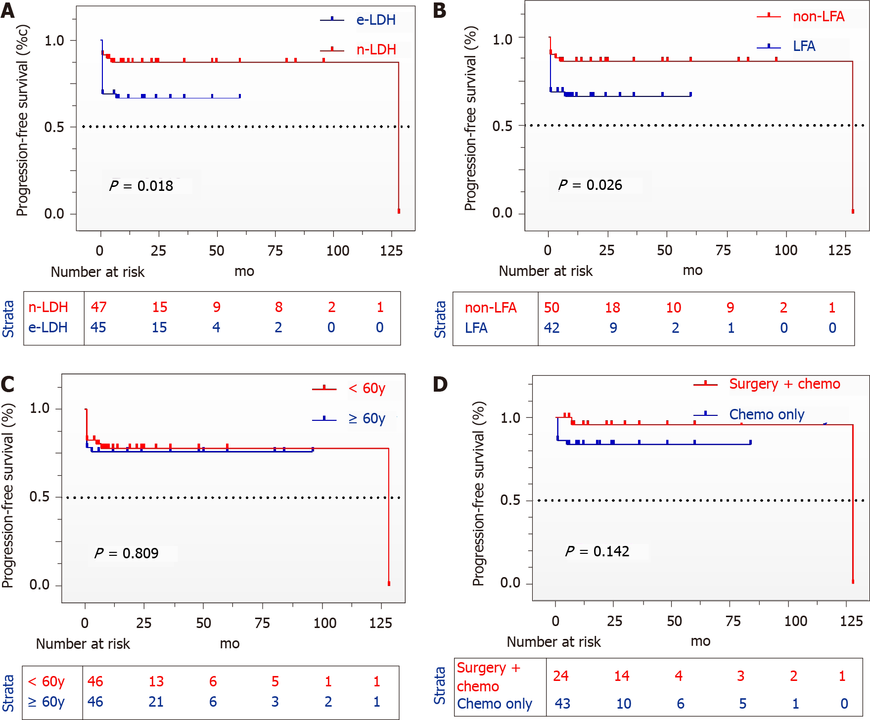
Figure 2 Progression-free-survival.
A: Kaplan-Meier plots showing the progression-free survival of patients with elevated lactate dehydrogenase (LDH) and normal LDH; B: Patients with normal/abnormal liver function; C: Patients with age less and over 60 years old; D: Patients with surgery plus chemotherapy and chemotherapy only. LDH: Lactate dehydrogenase; LFA: Liver function abnormality.
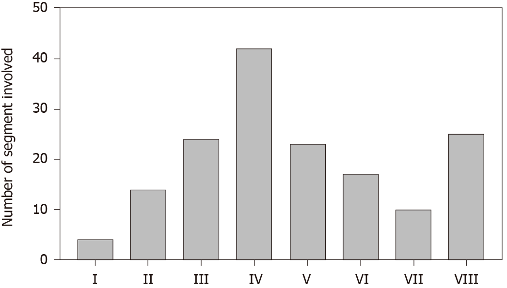
Figure 3 Number of segment involved.
A histogram of the liver segments afflicted by primary hepatic lymphoma presented in Rome numerals: segment I (4), II (14), III (24), IV (42), V (23), VI (17), VII (10), VIII (25).
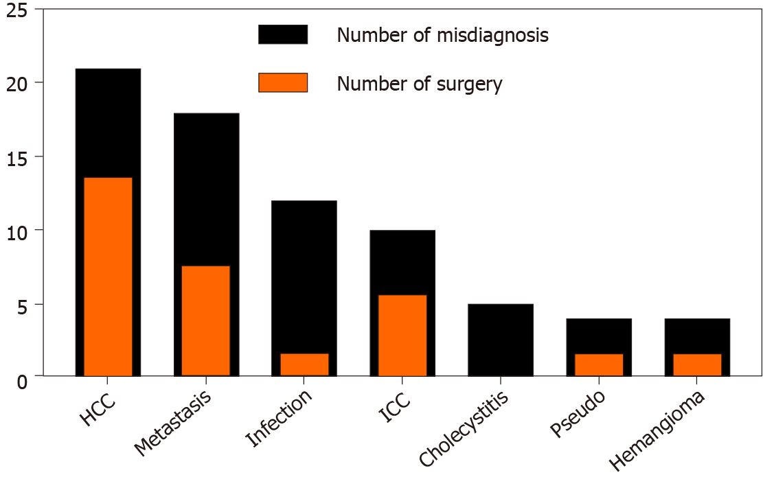
Figure 4 Number of misdiagnosis.
Classification of misdiagnosed primary hepatic lymphoma patients (black) and hepatectomized accordingly (orange). HCC: Hepatocellular carcinoma; ICC: Intrahepatic cholangiocarcinoma.
- Citation: Hai T, Zou LQ. Clinical management and susceptibility of primary hepatic lymphoma: A cases-based retrospective study. World J Clin Cases 2021; 9(31): 9417-9430
- URL: https://www.wjgnet.com/2307-8960/full/v9/i31/9417.htm
- DOI: https://dx.doi.org/10.12998/wjcc.v9.i31.9417


