Copyright
©The Author(s) 2021.
World J Clin Cases. Jan 26, 2021; 9(3): 722-735
Published online Jan 26, 2021. doi: 10.12998/wjcc.v9.i3.722
Published online Jan 26, 2021. doi: 10.12998/wjcc.v9.i3.722
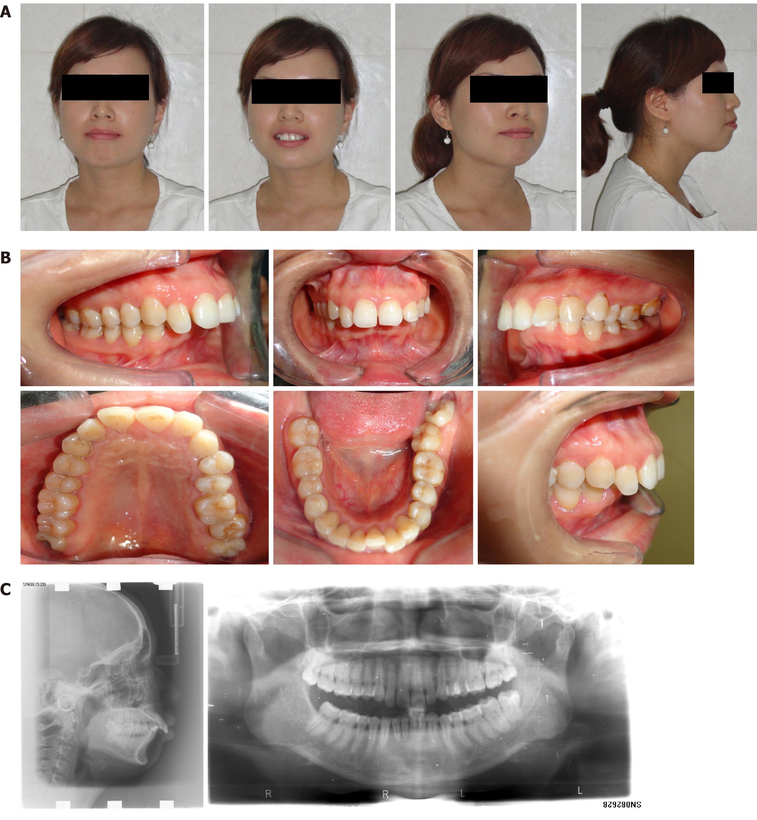
Figure 1 Pretreatment appearance.
A: Facial photographs; B: Intraoral photographs; C: Radiographs.
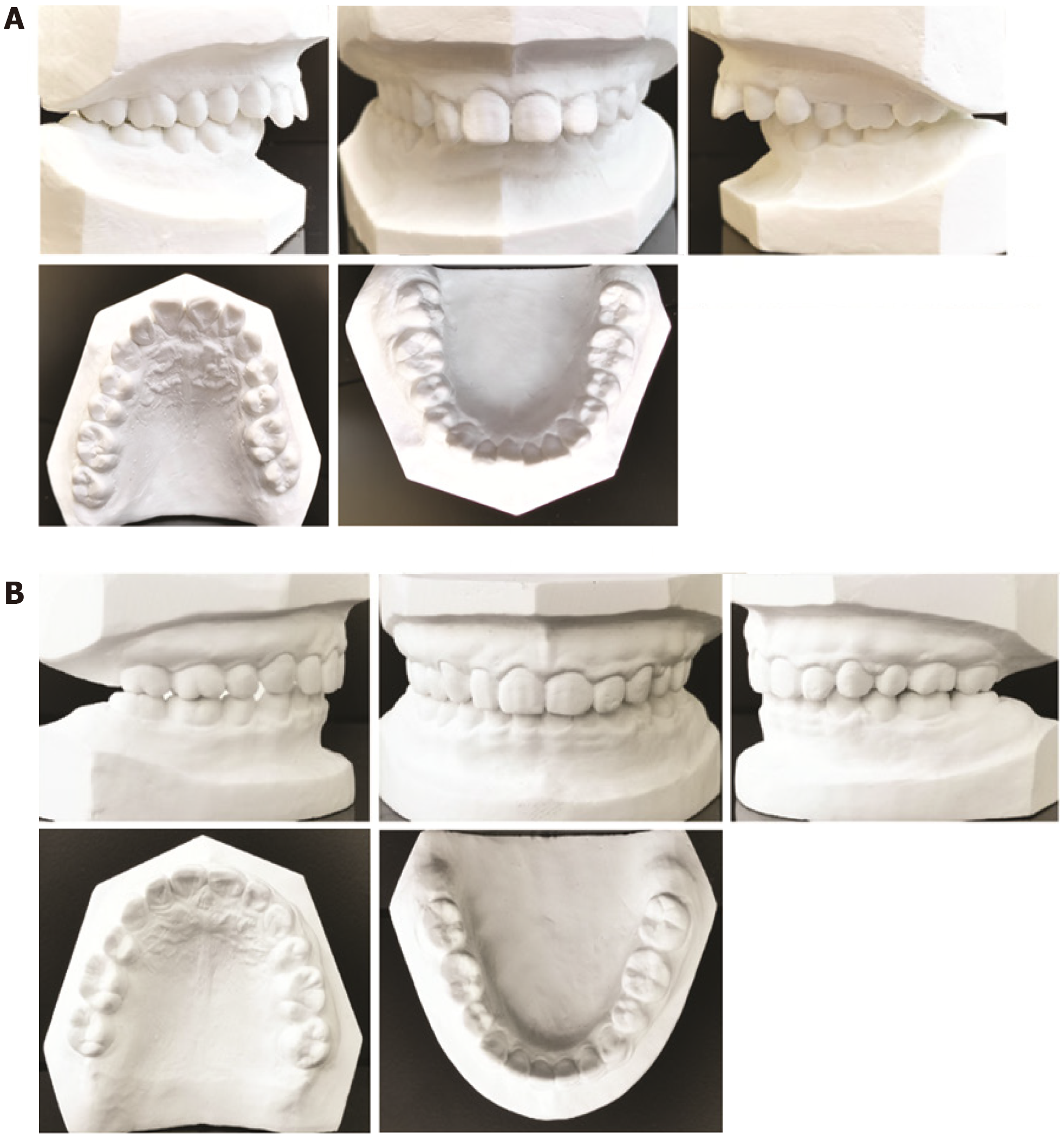
Figure 2 Dental casts.
A: Pretreatment; B: Posttreatment.
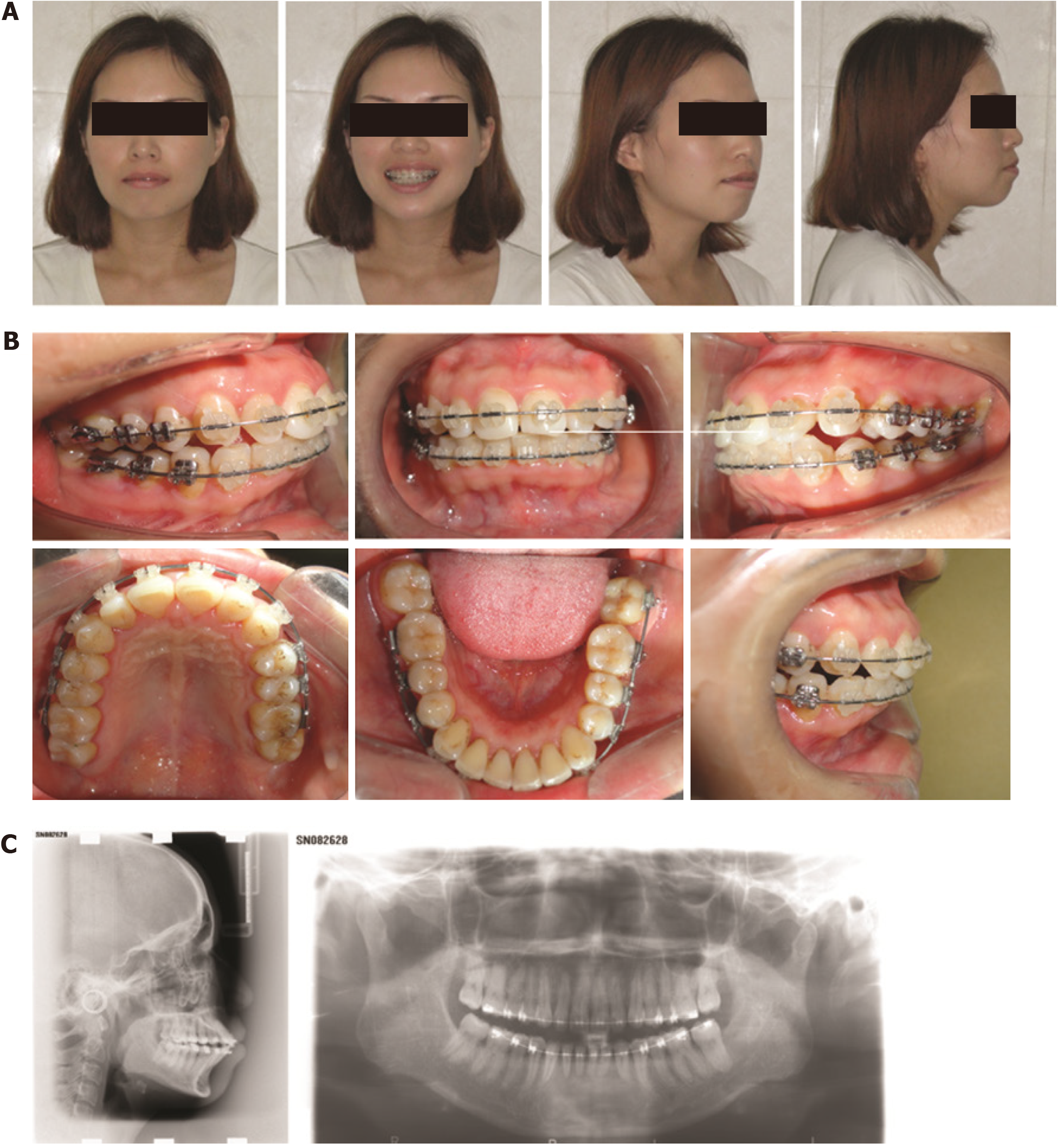
Figure 3 Appearance during treatment, after leveling and alignment.
A: Facial photographs; B: Intraoral photographs; C: Radiographs.
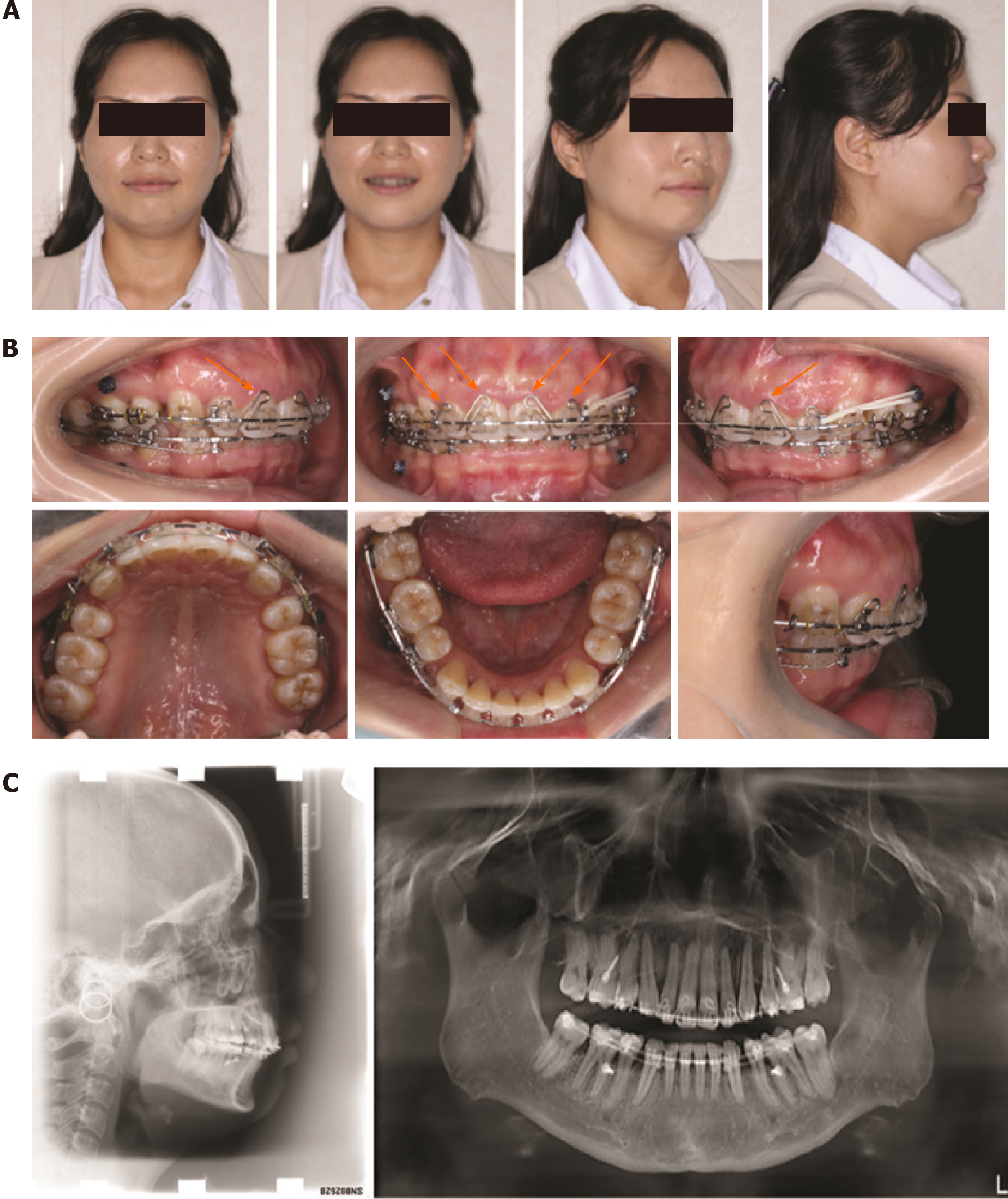
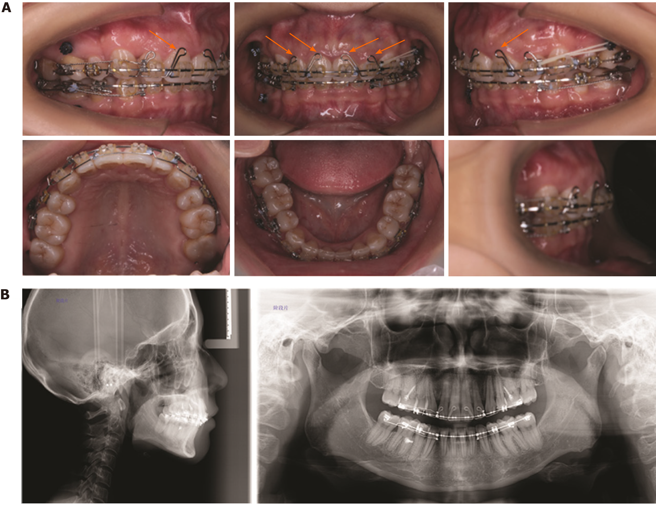
Figure 4 Appearance during treatment, at progress I stage after extraction of the four first premolars.
A: Facial photographs; B: Intraoral photographs (black arrows indicate the self-made four-curvature torquing auxiliary); C: Radiographs.
Figure 5 Appearance during treatment, at progress II stage after extraction of the four first premolars.
A: Intraoral photographs (black arrows indicate the self-made four-curvature torquing auxiliary); B: Radiographs.
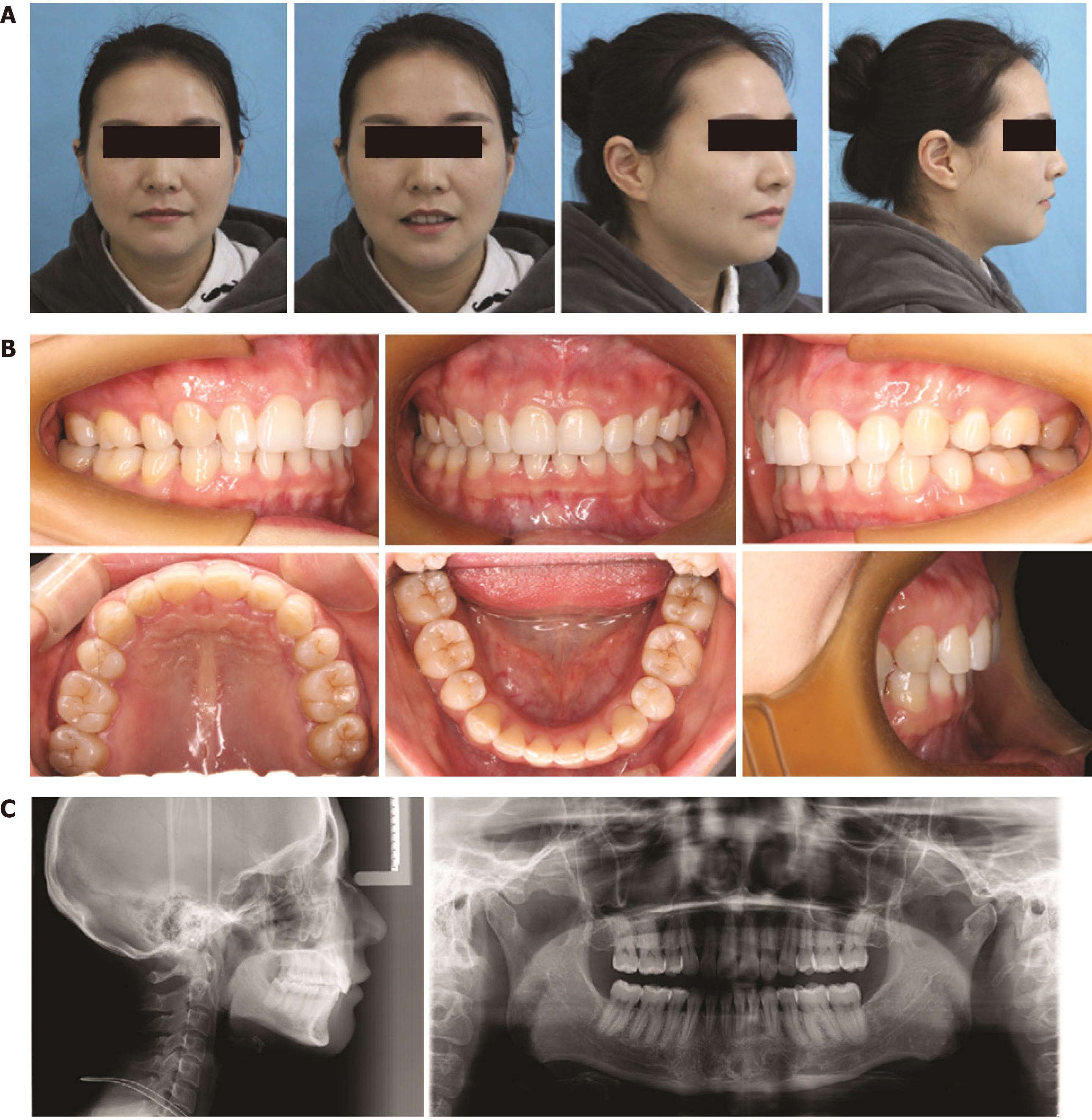
Figure 6 Posttreatment appearance.
A: Facial photographs; B: Intraoral photographs; C: Radiographs.
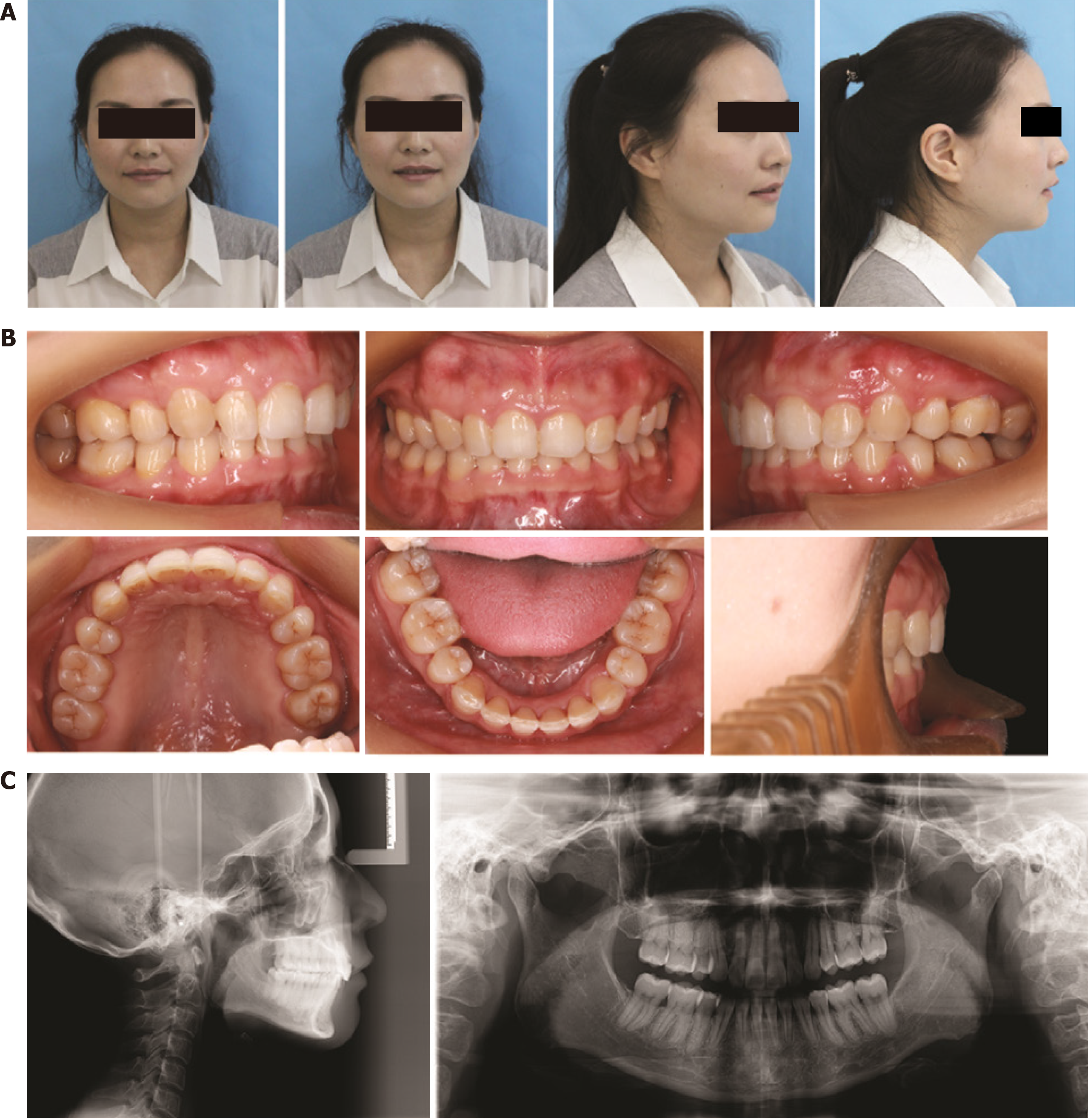
Figure 7 Appearance at 2-year follow-up.
A: Facial photographs; B: Intraoral photographs; C: Radiographs.
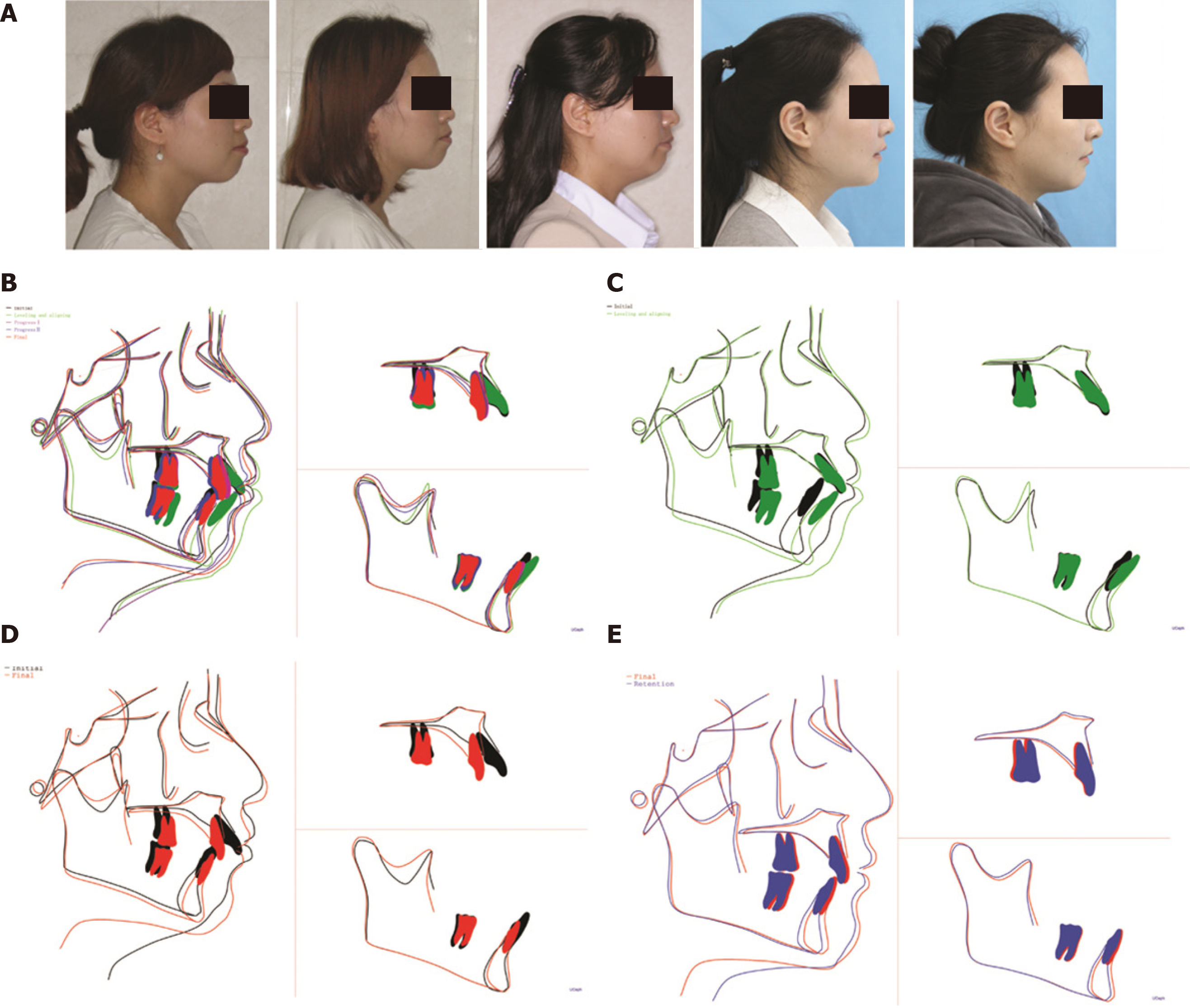
Figure 8 Facial profile photographs and cephalometric superimpositions.
A: Tracing comparison of facial profile photographs at the five different stages of pretreatment, leveling and aligning, progress I, posttreatment, and 2-year follow-up; B: Superimposition at the five stages; C: Superimposition at pretreatment (black) and leveling and aligning (green); D: Superimposition at pretreatment (black) and posttreatment (red); E: Superimposition at pretreatment (black) and 2-year follow-up (blue, showing retention).
- Citation: Liu R, Hou WB, Yang PZ, Zhu L, Zhou YQ, Yu X, Wen XJ. Severe skeletal bimaxillary protrusion treated with micro-implants and a self-made four-curvature torquing auxiliary: A case report. World J Clin Cases 2021; 9(3): 722-735
- URL: https://www.wjgnet.com/2307-8960/full/v9/i3/722.htm
- DOI: https://dx.doi.org/10.12998/wjcc.v9.i3.722


