Copyright
©The Author(s) 2021.
World J Clin Cases. Jan 26, 2021; 9(3): 714-721
Published online Jan 26, 2021. doi: 10.12998/wjcc.v9.i3.714
Published online Jan 26, 2021. doi: 10.12998/wjcc.v9.i3.714
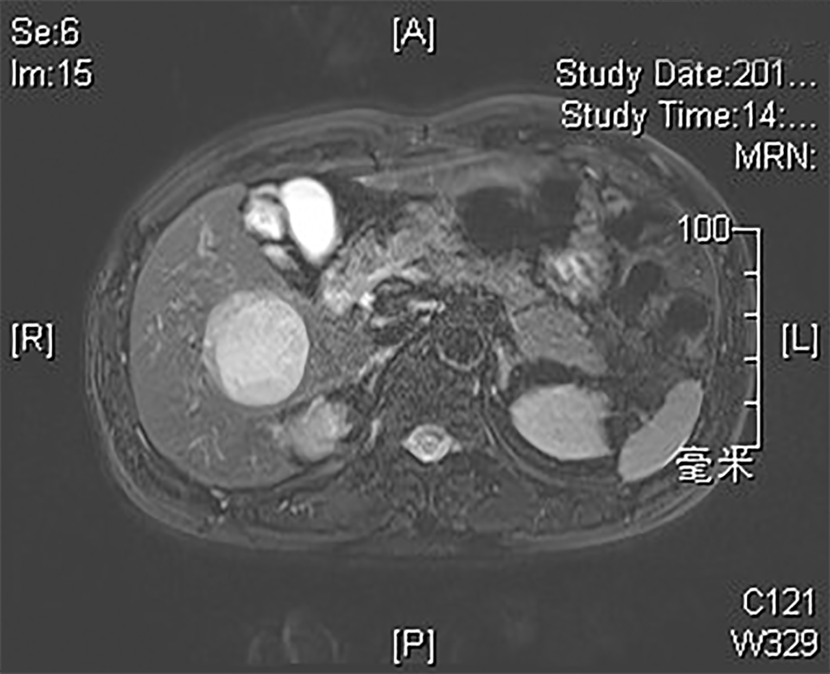
Figure 1 Location map of the liver tumor.
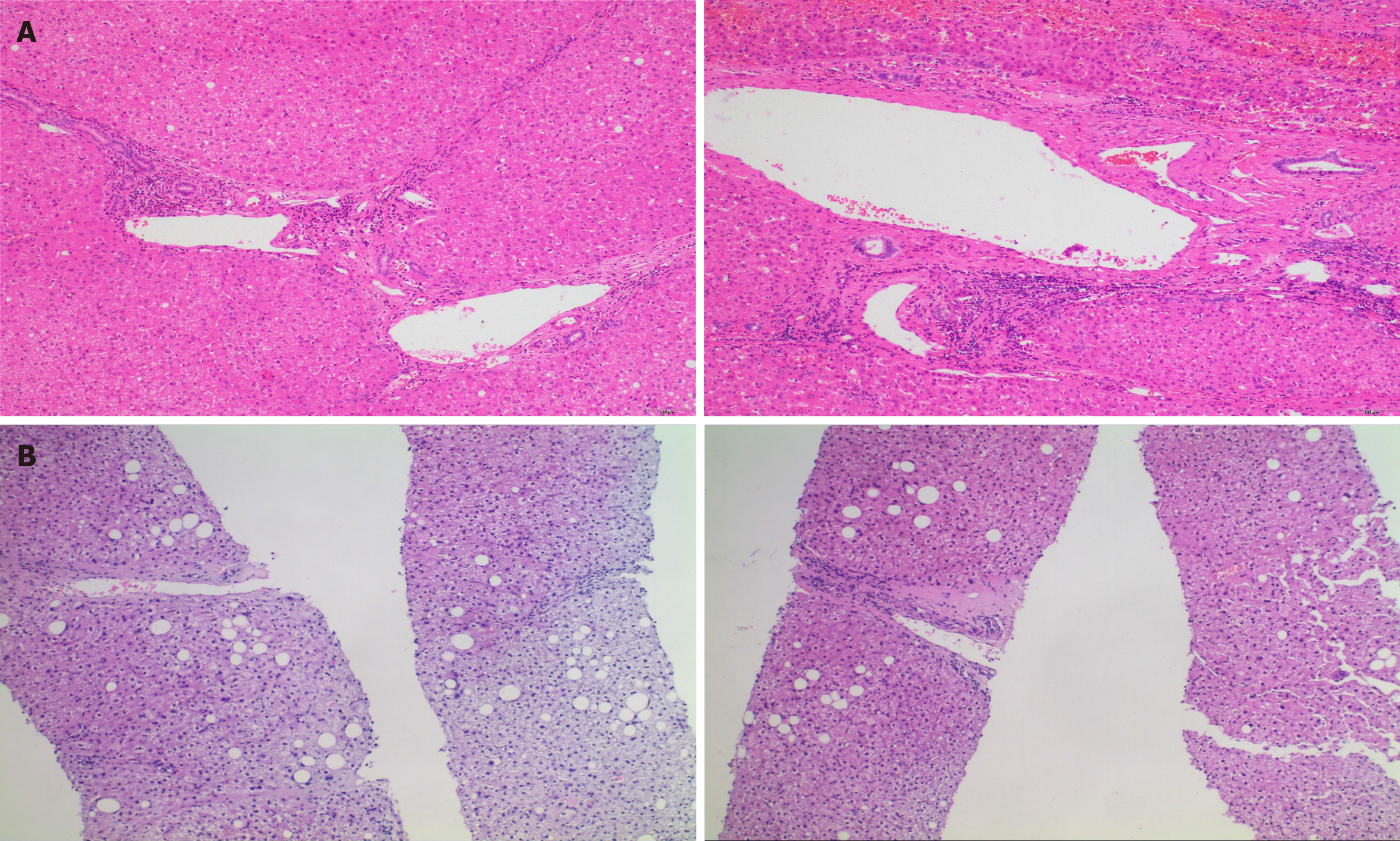
Figure 2 Histology.
A: Pathological staining of the resected liver cancer tissue at Zhongshan Hospital, Shanghai in October 2016 (G1S3); B: Liver puncture biopsy (G2S1) performed at Quanzhou First Hospital in July 2020.
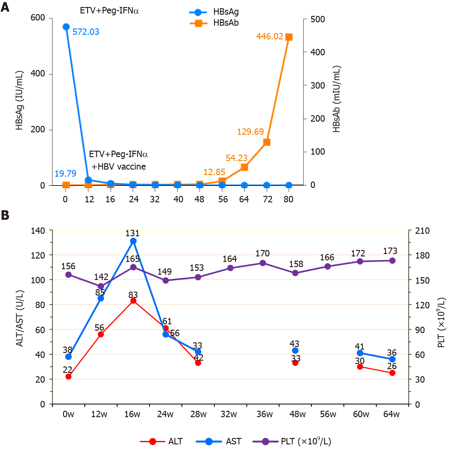
Figure 3 Changes in the laboratory examination results during treatment.
A: Changes in the hepatitis B virology indices HBsAg and HBsAb; B: Changes in alanine aminotransferase and aspartate aminotransferase levels, and platelet count. ETV: Entecavir; Peg-IFNα: Pegylated interferon alpha-2b; HBV: Hepatitis B virus; AST: Alanine aminotransferase; ALT: Aspartate aminotransferase; PLT: Platelet count.
- Citation: Yu XP, Lin Q, Huang ZP, Chen WS, Zheng MH, Zheng YJ, Li JL, Su ZJ. Clinical cure and liver fibrosis reversal after postoperative antiviral combination therapy in hepatitis B-associated non-cirrhotic hepatocellular carcinoma: A case report. World J Clin Cases 2021; 9(3): 714-721
- URL: https://www.wjgnet.com/2307-8960/full/v9/i3/714.htm
- DOI: https://dx.doi.org/10.12998/wjcc.v9.i3.714


