Copyright
©The Author(s) 2021.
World J Clin Cases. Jan 26, 2021; 9(3): 707-713
Published online Jan 26, 2021. doi: 10.12998/wjcc.v9.i3.707
Published online Jan 26, 2021. doi: 10.12998/wjcc.v9.i3.707
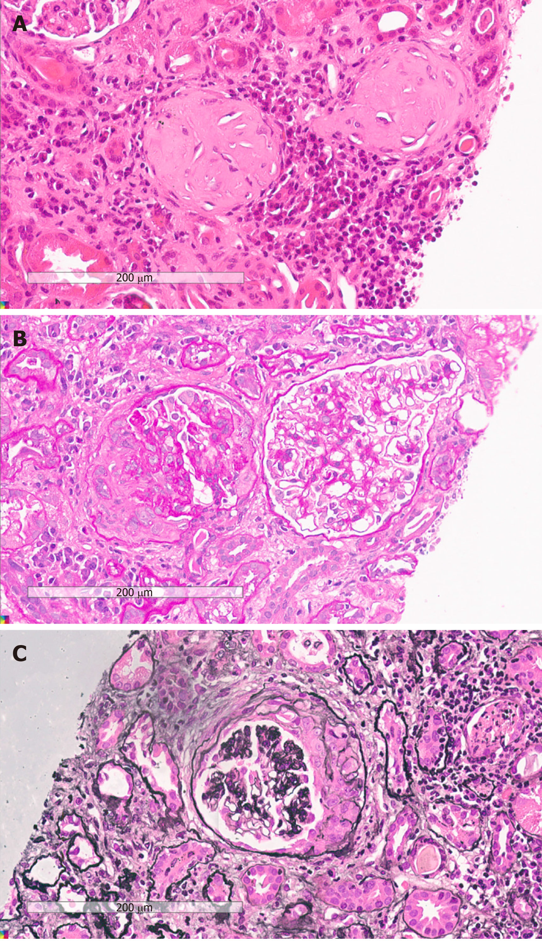
Figure 1 Sections of kidney samples taken at autopsy showing sclerotic glomerulus, fibro-cellular crescent, and cellular crescent.
A: Haematoxylin and eosin stained image; B: Periodic acid-Schiff reaction; C: Periodic acid-Schiff-methenamine silver staining. Original magnification × 400.
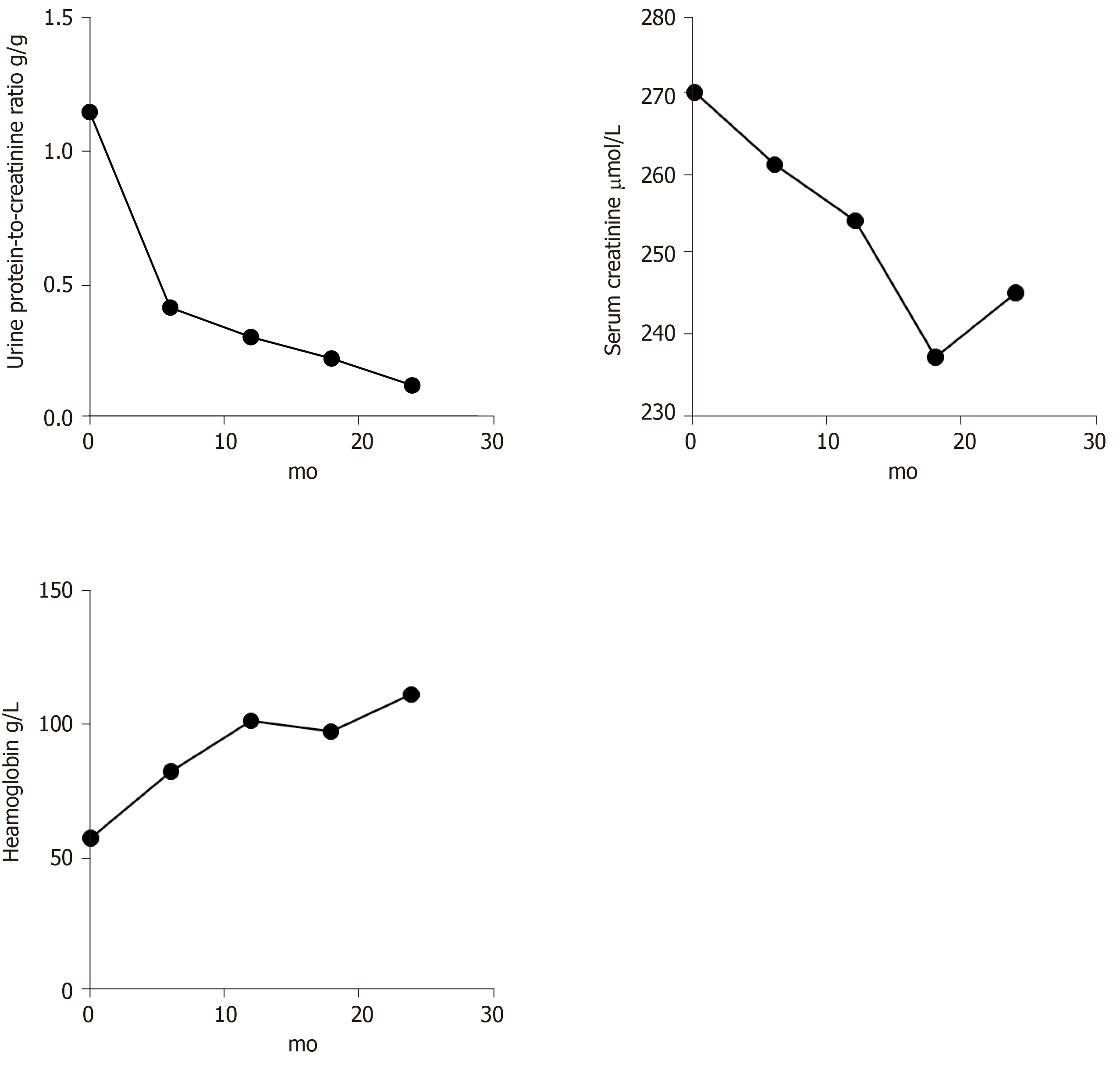
Figure 2 Trends of urine protein-to-creatinine ratio, serum creatinine, and haemoglobin.
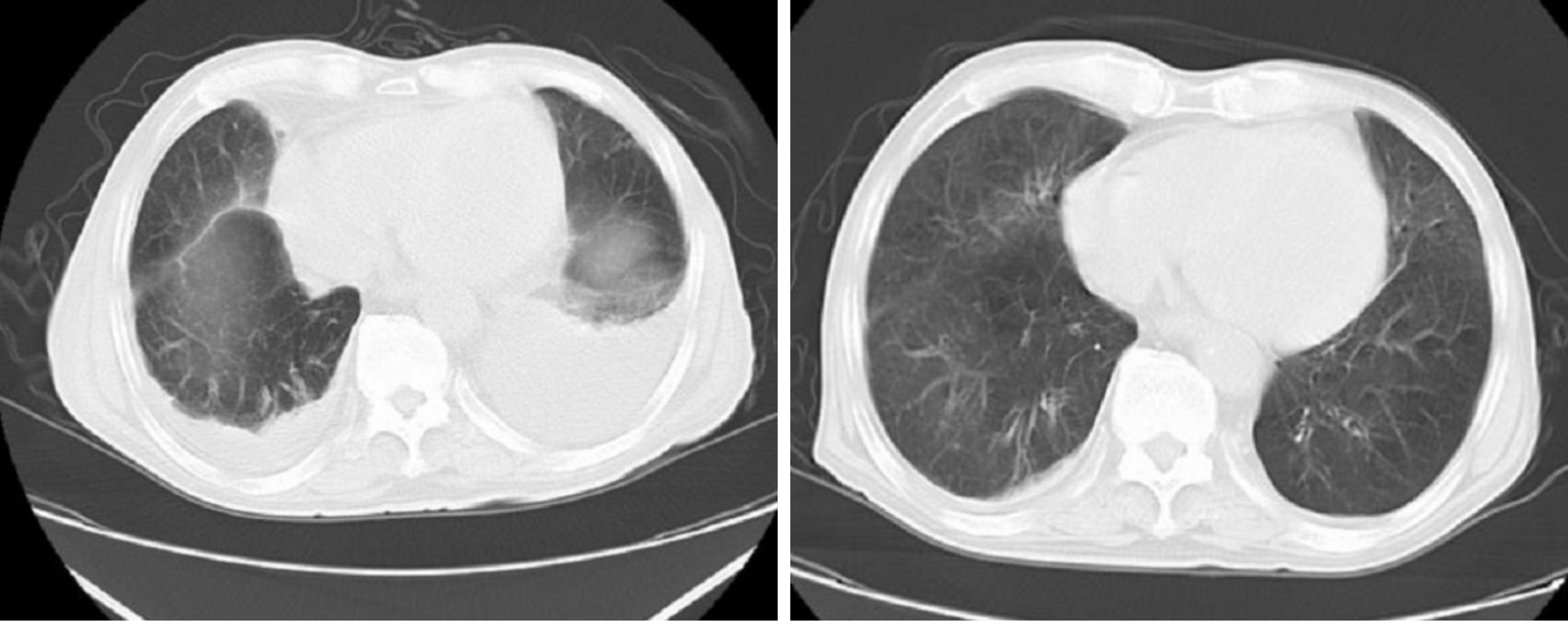
Figure 3 Computed tomography of the chest showing pleural effusion.
- Citation: Xu ZG, Li WL, Wang X, Zhang SY, Zhang YW, Wei X, Li CD, Zeng P, Luan SD. Systemic lupus erythematosus and antineutrophil cytoplasmic antibody-associated vasculitis overlap syndrome in a 77-year-old man: A case report. World J Clin Cases 2021; 9(3): 707-713
- URL: https://www.wjgnet.com/2307-8960/full/v9/i3/707.htm
- DOI: https://dx.doi.org/10.12998/wjcc.v9.i3.707


