Copyright
©The Author(s) 2021.
World J Clin Cases. Jan 26, 2021; 9(3): 690-696
Published online Jan 26, 2021. doi: 10.12998/wjcc.v9.i3.690
Published online Jan 26, 2021. doi: 10.12998/wjcc.v9.i3.690
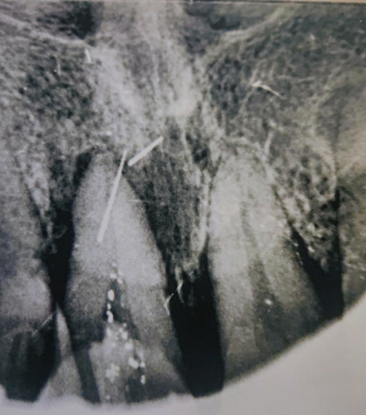
Figure 1 Dental film showing the foreign body at position 21.
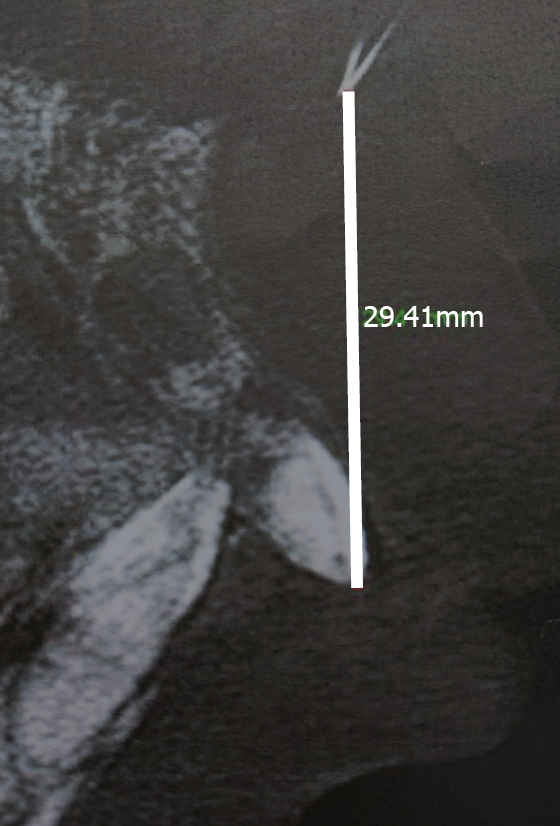
Figure 2 Postoperative dental film showing the foreign body residue 29.
4 mm from the tip of the left incisor.
Figure 3 Computed tomography scan of the nasal sinuses before the third surgery.
A: Axial plane; B: Sagittal plane; C: Coronal plane showing a pin-like foreign body with hyperdensity in the left side of the nasal septum; D: Three-dimensional reconstruction reconfirmed the position.
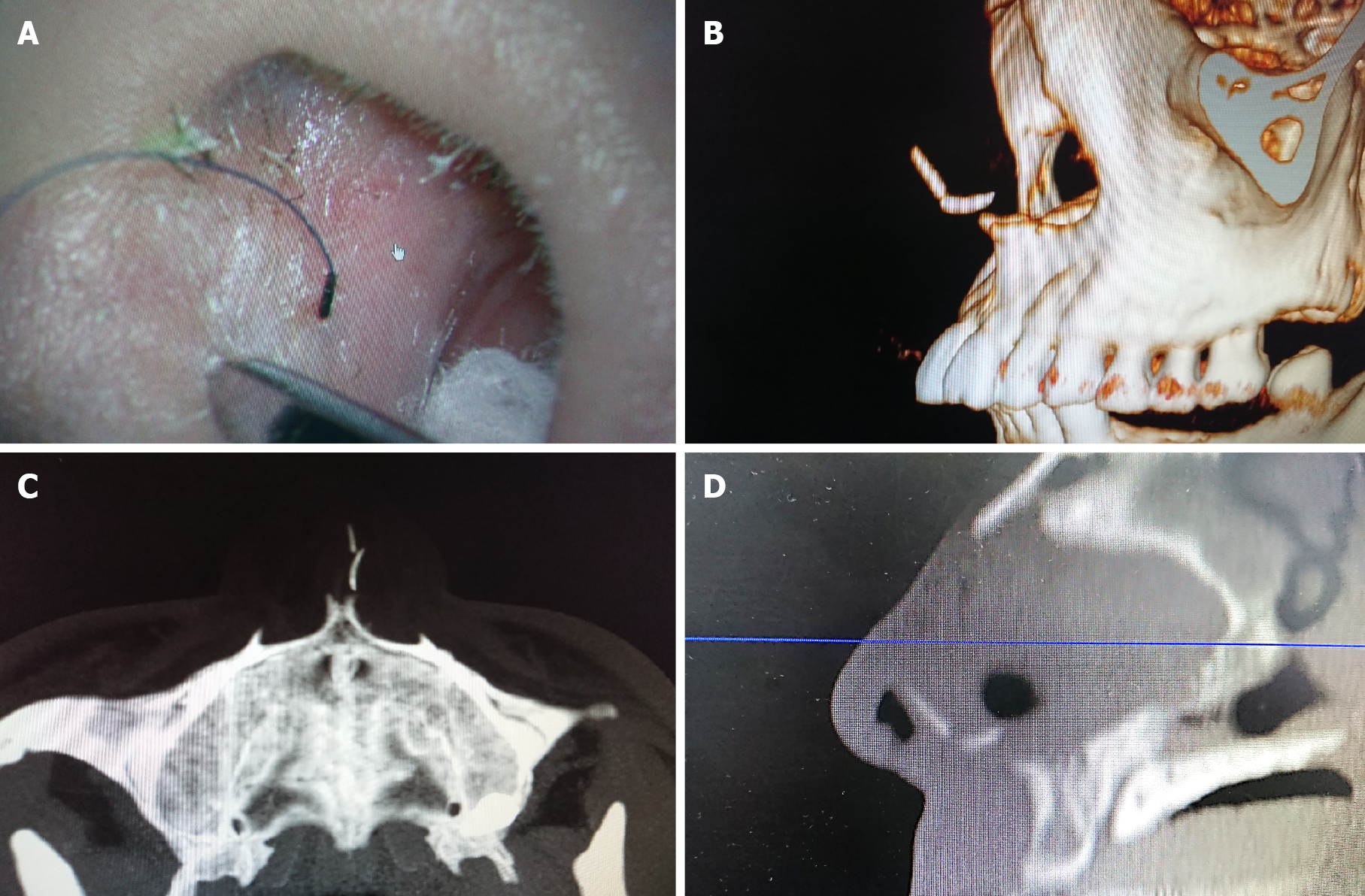
Figure 4 Computed tomography scan of the nasal sinuses after the third surgery.
A: A needle was used as a reference; B: Three-dimensional reconstruction; C and D: Axial (C) and sagittal planes (D) showing that the needle was pointing right to the pin-like foreign body.
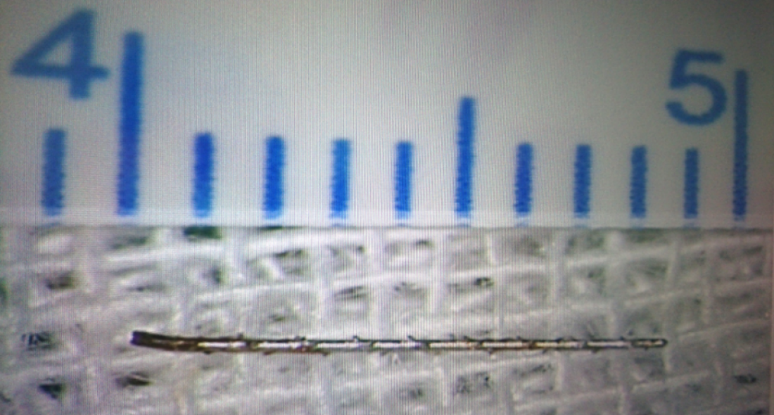
Figure 5 The gross of the foreign body confirmed to be a piece of the fractured instrument for root canal therapy.
- Citation: Du XW, Zhang JB, Xiao SF. Nasal septal foreign body as a complication of dental root canal therapy: A case report. World J Clin Cases 2021; 9(3): 690-696
- URL: https://www.wjgnet.com/2307-8960/full/v9/i3/690.htm
- DOI: https://dx.doi.org/10.12998/wjcc.v9.i3.690


