Copyright
©The Author(s) 2021.
World J Clin Cases. Jan 26, 2021; 9(3): 666-671
Published online Jan 26, 2021. doi: 10.12998/wjcc.v9.i3.666
Published online Jan 26, 2021. doi: 10.12998/wjcc.v9.i3.666
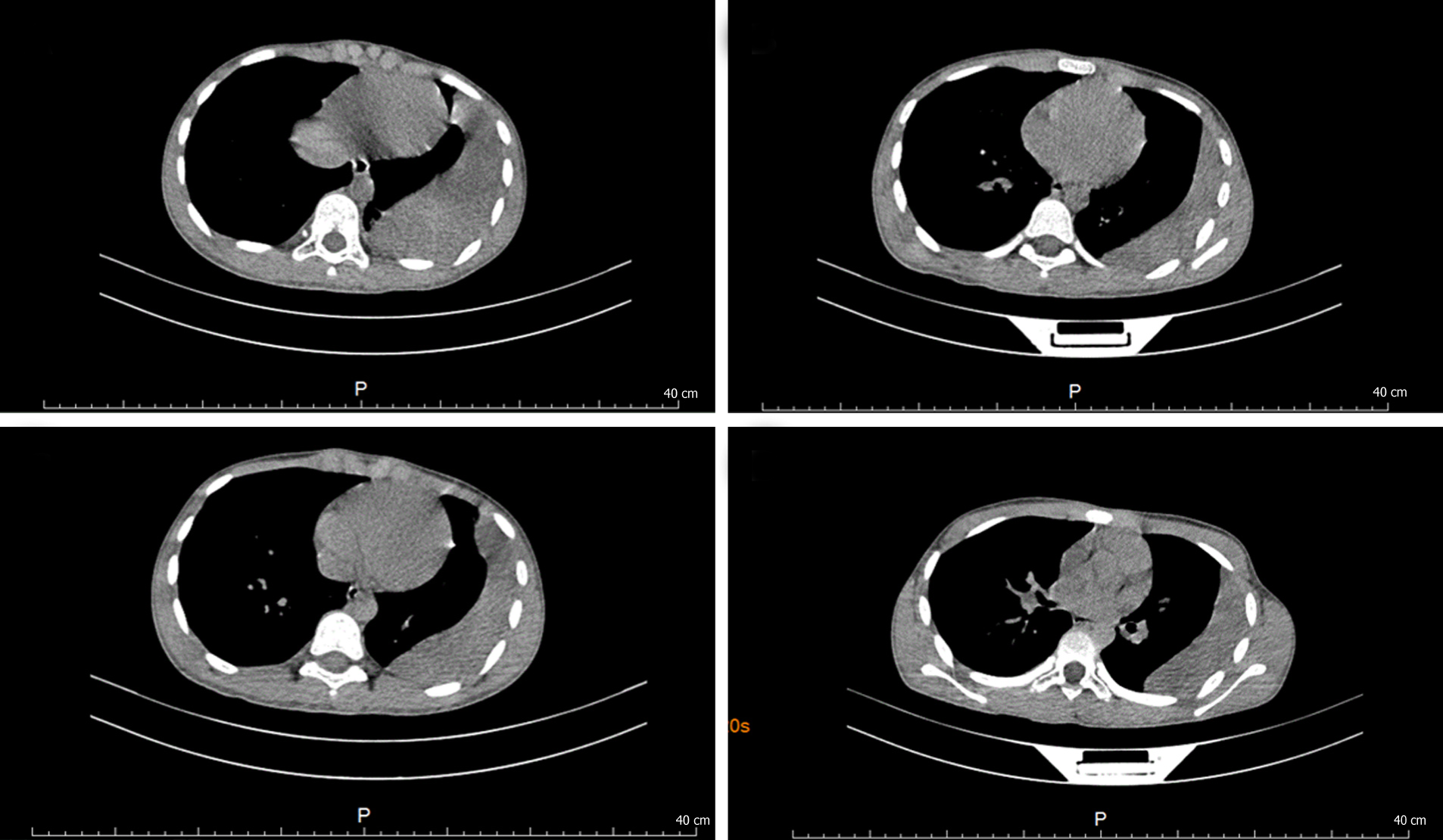
Figure 1 Computed tomography scan of the patient before the praziquantel treatment.
Moderate effusion in the left pleural cavity, left pleural thickening and calcification, and a small amount of patchy and striped shadows was visible on the left lower and upper lobes.
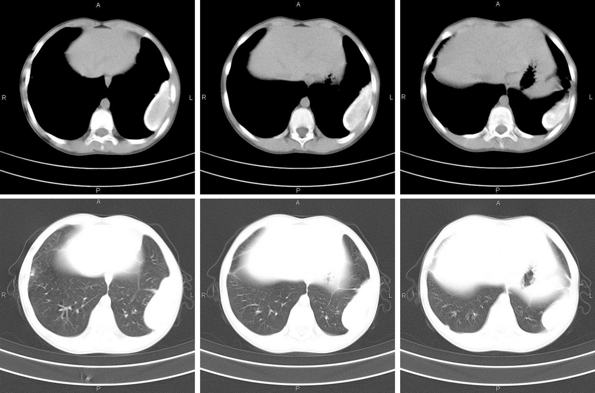
Figure 2 Computed tomography scan of the patient several months after the praziquantel treatment.
An uneven hyperdensity cystic nodule with smooth edges was seen on the left inferolateral lobe. The largest transverse section was about 6.0 cm × 2.3 cm, with no obvious enhancement inside; the margin of the nodule calcified and was adhesive to the chest wall, and the corresponding pleural thickened.
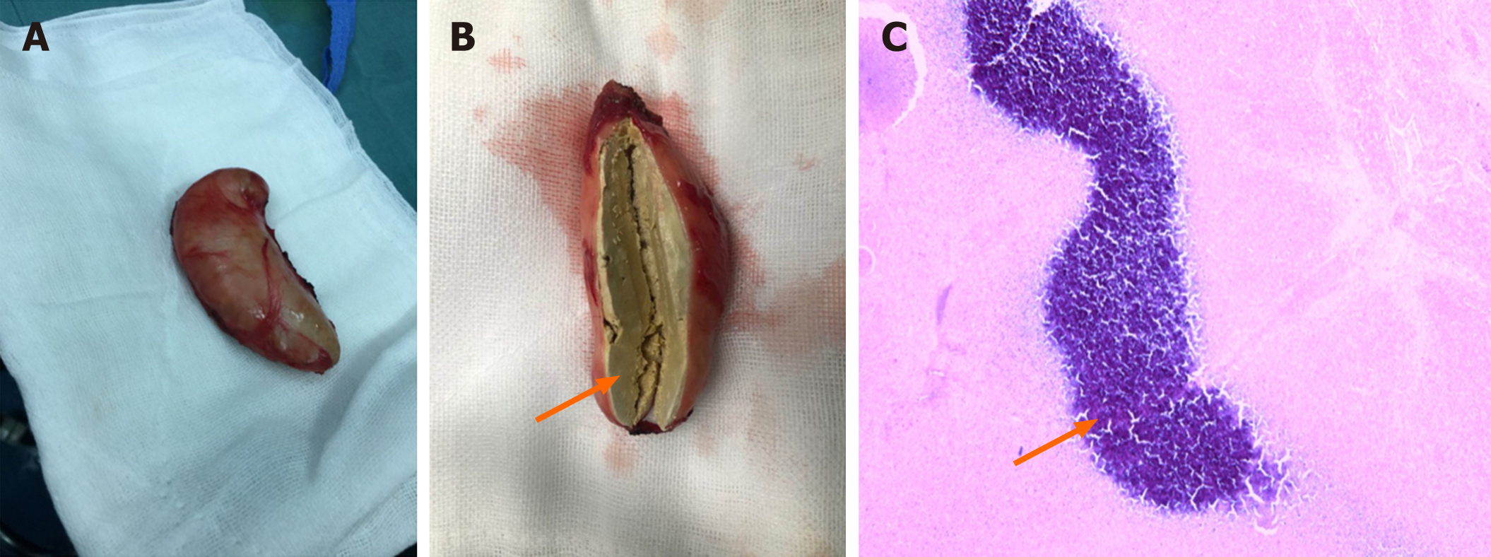
Figure 3 Gross specimen and biopsy of the resected mass.
A: The gross specimen of the mass showing an encapsulated nodule; B: The cut surface of the mass. The arrowhead indicates the necrotic tissue inside; C: The biopsy of the mass. The cystic wall of the mass consists of fibrous tissue. The blue-stained area indicated by the arrowhead is non-structured necrotic tissue.
- Citation: Xie Y, Luo YR, Chen M, Xie YM, Sun CY, Chen Q. Pleural lump after paragonimiasis treated by thoracoscopy: A case report. World J Clin Cases 2021; 9(3): 666-671
- URL: https://www.wjgnet.com/2307-8960/full/v9/i3/666.htm
- DOI: https://dx.doi.org/10.12998/wjcc.v9.i3.666


