Copyright
©The Author(s) 2021.
World J Clin Cases. Oct 6, 2021; 9(28): 8595-8601
Published online Oct 6, 2021. doi: 10.12998/wjcc.v9.i28.8595
Published online Oct 6, 2021. doi: 10.12998/wjcc.v9.i28.8595
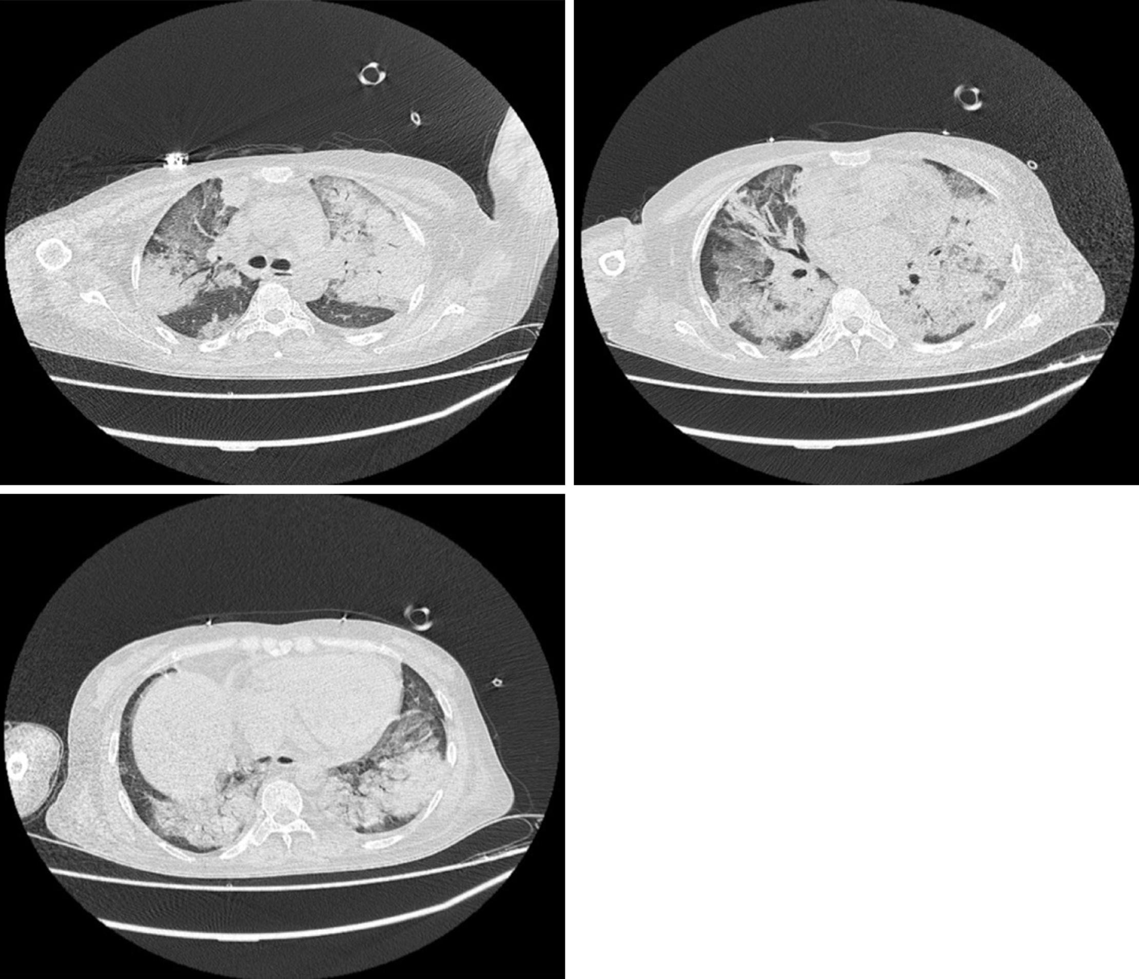
Figure 1 Chest computed tomography indicated multiple consolidation shadows and plaques in both lungs, and pulmonary edema with inflammation was considered.
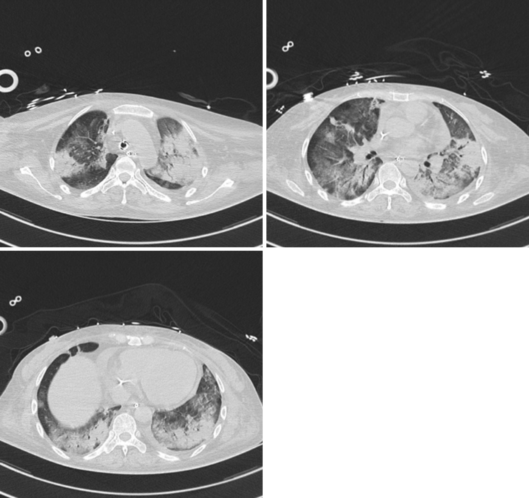
Figure 2 After anti-infection treatment, the consolidation of the lung disappeared slightly.
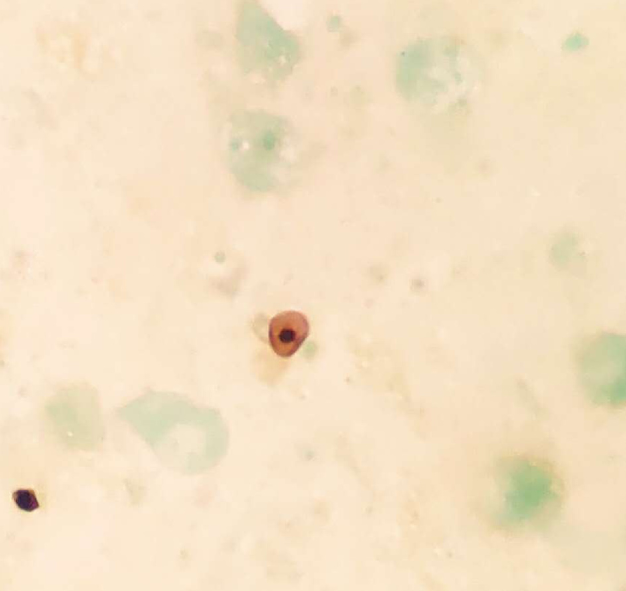
Figure 3 Pneumocystis was found in the patient's bronchoalveolar lavage fluid by hexamine silver staining.
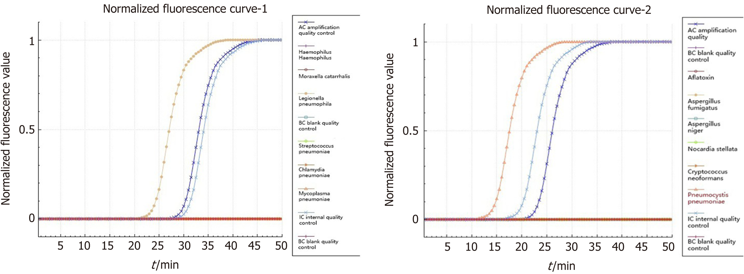
Figure 4 Isothermal chip amplification were used to verify Legionella and Pneumocystis.
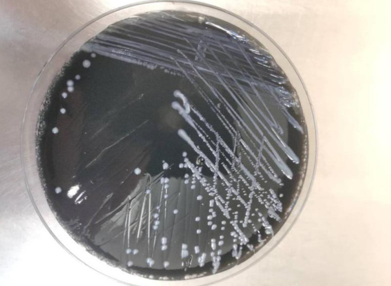
Figure 5 Legionella colonies on BCYE medium.
- Citation: Wu WH, Hui TC, Wu QQ, Xu CA, Zhou ZW, Wang SH, Zheng W, Yin QQ, Li X, Pan HY. Pneumocystis jirovecii and Legionella pneumophila coinfection in a patient with diffuse large B-cell lymphoma: A case report. World J Clin Cases 2021; 9(28): 8595-8601
- URL: https://www.wjgnet.com/2307-8960/full/v9/i28/8595.htm
- DOI: https://dx.doi.org/10.12998/wjcc.v9.i28.8595


