Copyright
©The Author(s) 2021.
World J Clin Cases. Oct 6, 2021; 9(28): 8587-8594
Published online Oct 6, 2021. doi: 10.12998/wjcc.v9.i28.8587
Published online Oct 6, 2021. doi: 10.12998/wjcc.v9.i28.8587
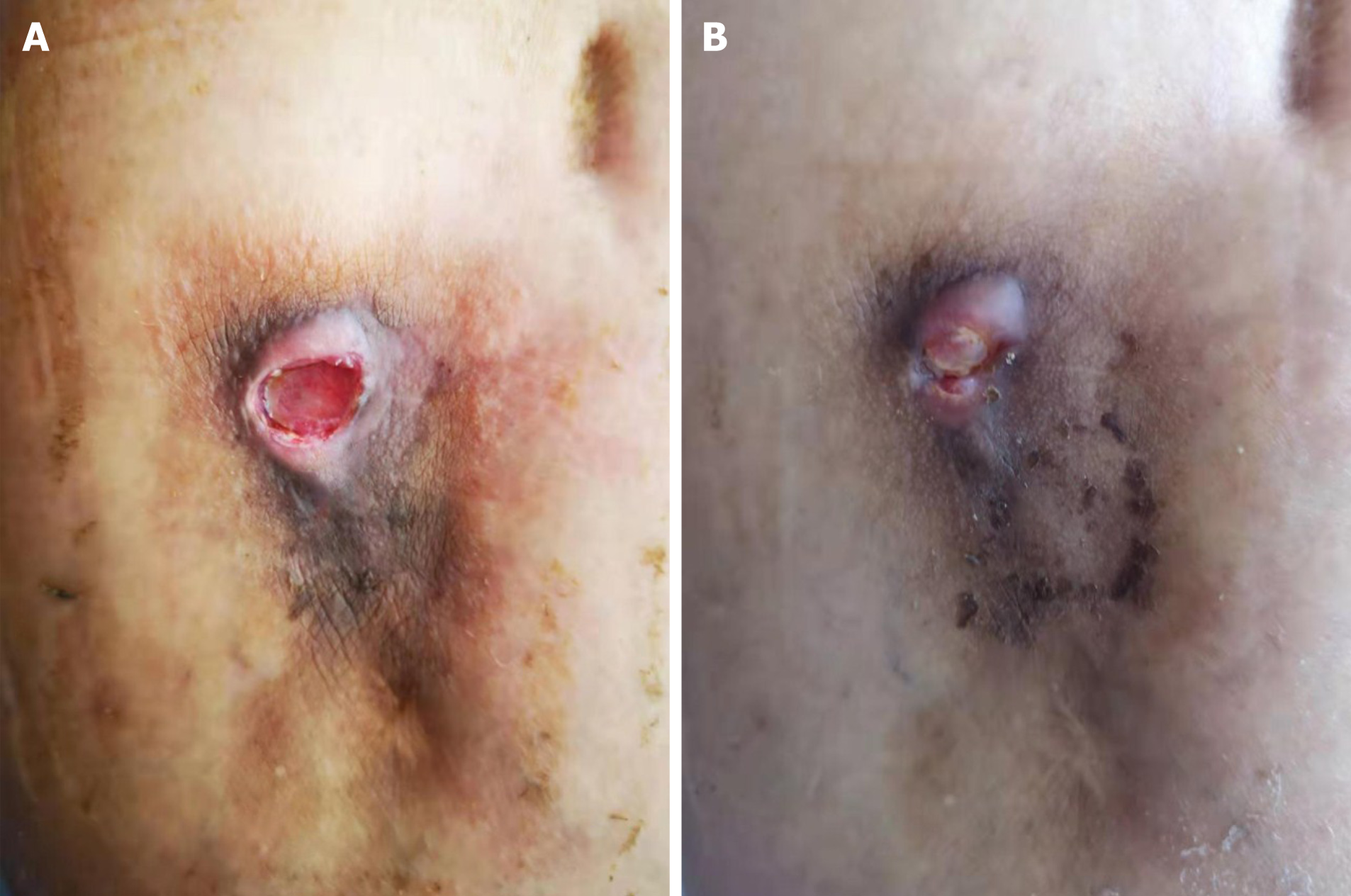
Figure 1 Performance of the drainage sinus.
A: There was plenty of fresh granulation tissue in a drainage sinus; B: After 22 d of anti-tuberculosis chemotherapy treatment, the sinus was almost healed.
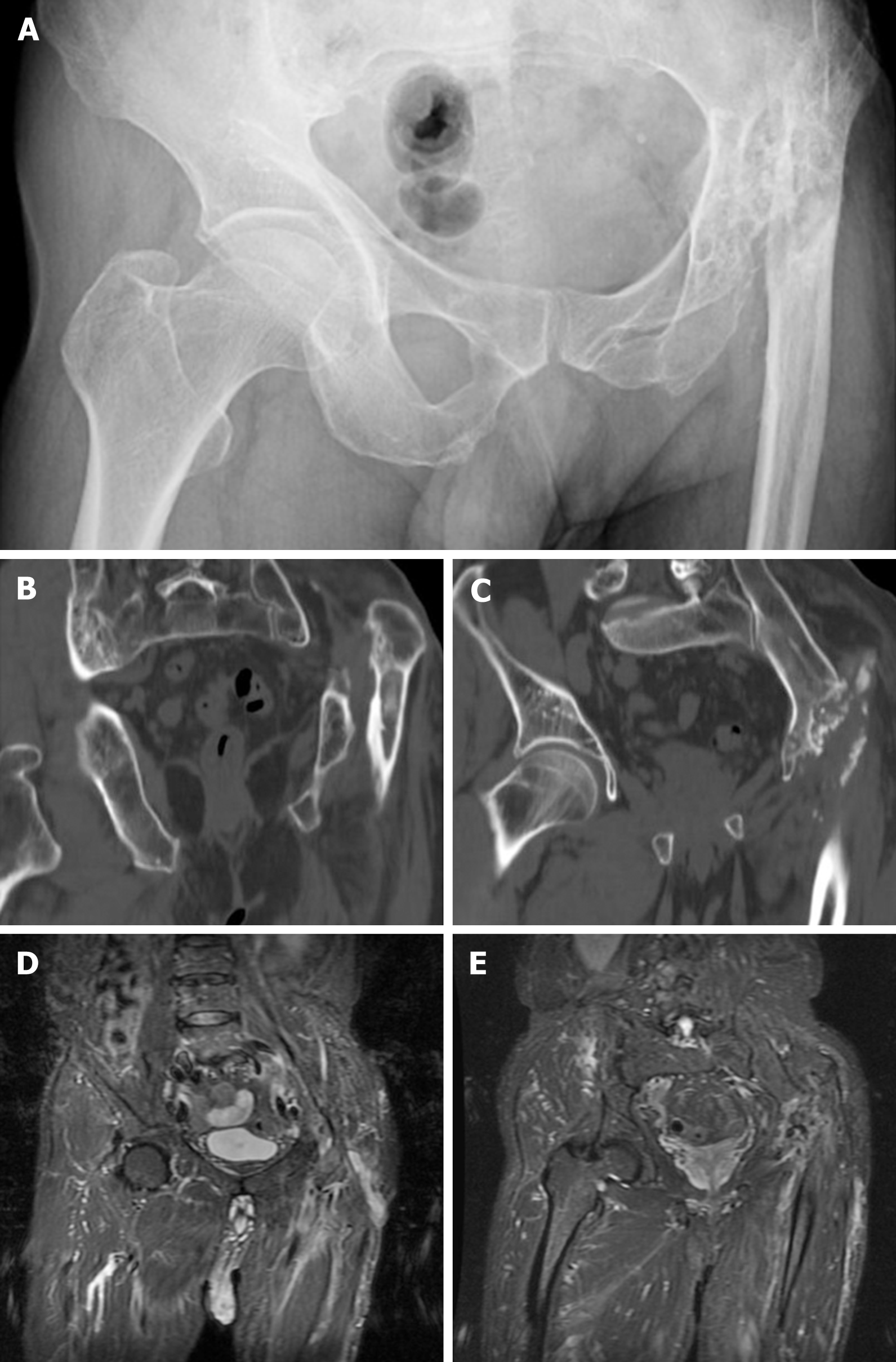
Figure 2 Preoperative imaging finding.
A: The preoperative X-ray showed heavy destruction of the left femoral head and neck, pseudarthrosis between the proximal femur and iliac ala, and a smaller and shallower original acetabulum; B and C: The preoperative computed tomography showed heavy destruction of the left femoral head and neck, pseudarthrosis between the proximal femur and iliac ala, and a smaller and shallower original acetabulum; D and E: The preoperative magnetic resonance showed a slender form localized sinus and no infective foci in the pelvic cavity.
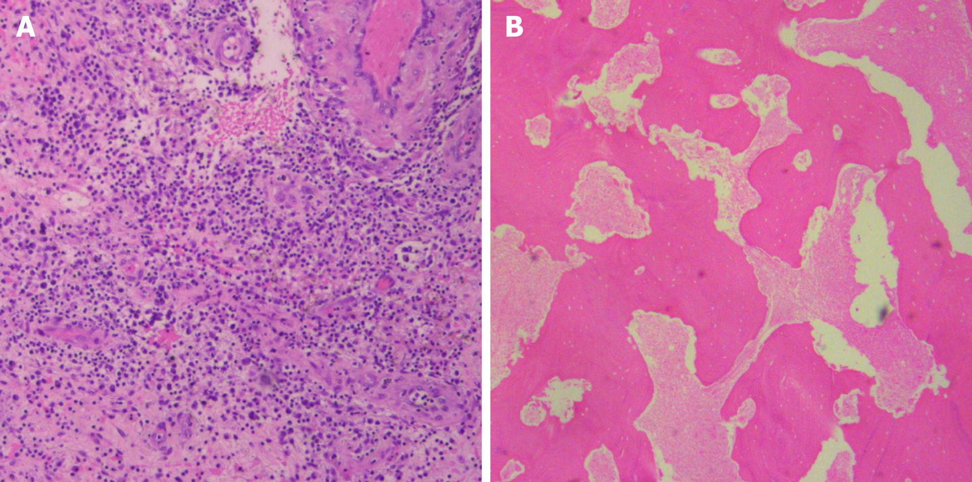
Figure 3 Intraoperative rapid pathology and postoperative pathology.
A: The intraoperative rapid pathological diagnosis showed no apparent active infection; B: The pathological examination of lesion tissue showed evident caseous necrosis.
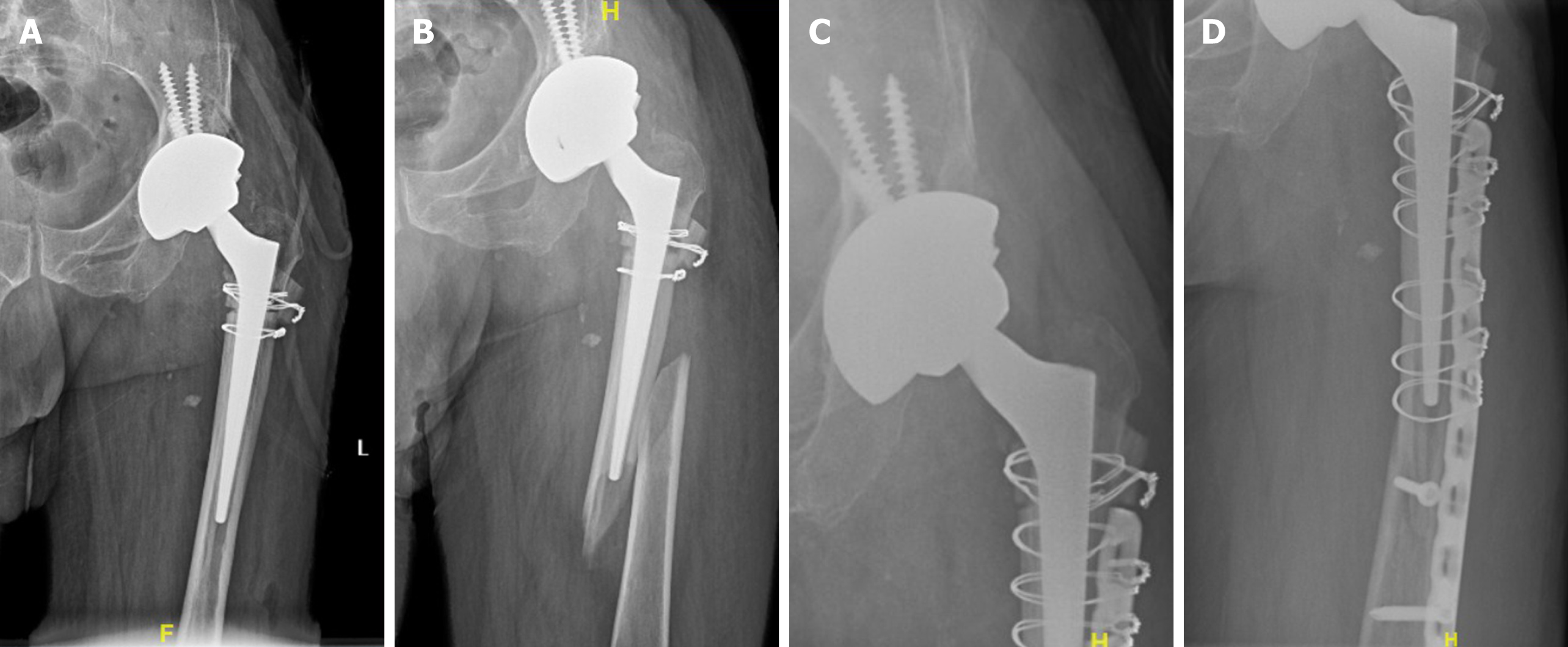
Figure 4 Postoperative imaging findings.
A: The postoperative X-ray showed that the prosthesis was in a good position; B: The X-ray showed periprosthetic fractures, no loss of the prostheses, and no destruction of the proximal femur; C and D: Three months after internal fixation, the X-ray of the left hip showed no recurrent bone destruction or prosthesis loss.
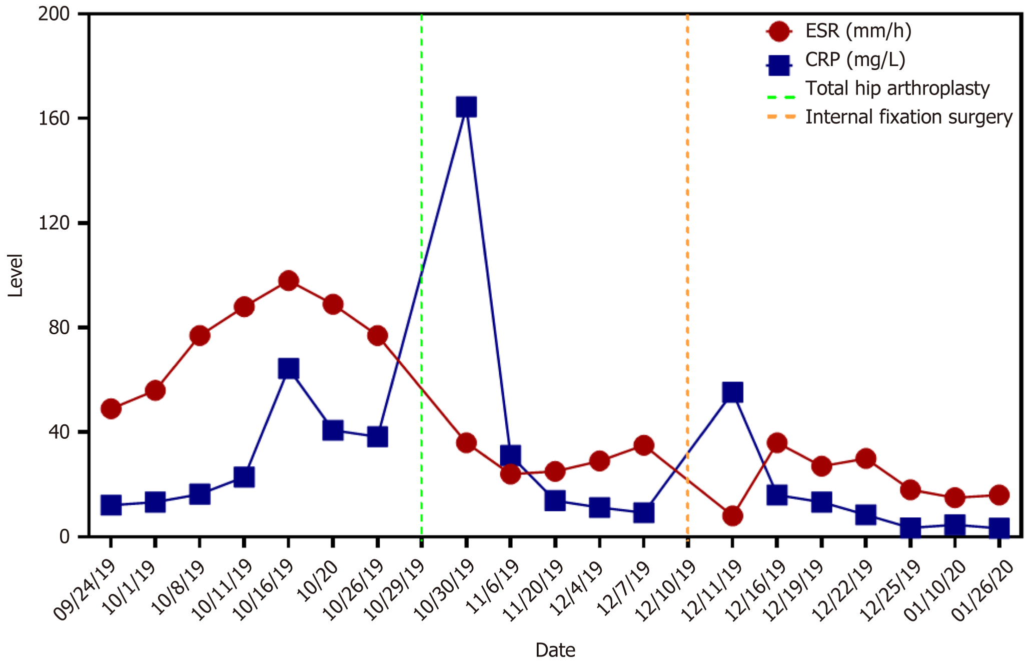
Figure 5 The trends of erythrocyte sedimentation rate and a C-reactive protein level showed that erythrocyte sedimentation rate and C-reactive protein returned to normal after 89 d of anti-tuberculosis chemotherapy after one-stage total hip arthroplasty.
In addition, the tests were negative three times in a row. ESR: Erythrocyte sedimentation rate; CRP: C-reactive protein.
- Citation: Zhu RT, Shen LP, Chen LL, Jin G, Jiang HT. One-stage total hip arthroplasty for advanced hip tuberculosis combined with developmental dysplasia of the hip: A case report. World J Clin Cases 2021; 9(28): 8587-8594
- URL: https://www.wjgnet.com/2307-8960/full/v9/i28/8587.htm
- DOI: https://dx.doi.org/10.12998/wjcc.v9.i28.8587


