Copyright
©The Author(s) 2021.
World J Clin Cases. Oct 6, 2021; 9(28): 8545-8551
Published online Oct 6, 2021. doi: 10.12998/wjcc.v9.i28.8545
Published online Oct 6, 2021. doi: 10.12998/wjcc.v9.i28.8545
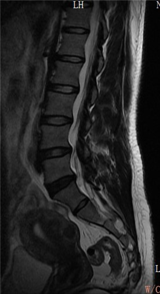
Figure 1 Lumbar spine magnetic resonance imaging indicated degenerative changes in the L5-S1 disc and with no sign of spinal nerve compression.
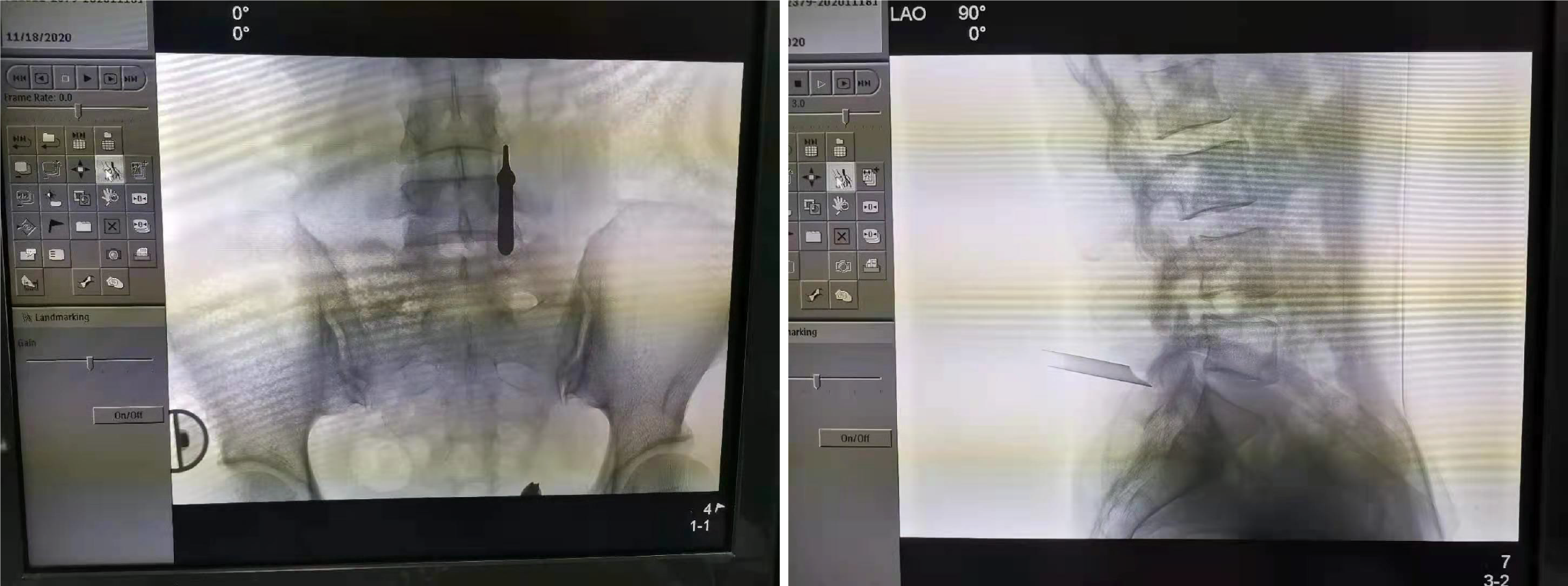
Figure 2 Anterior-posterior view and lateral view confirmed that the working cannula located on the posterior surface of the L5-S1 facet joint.
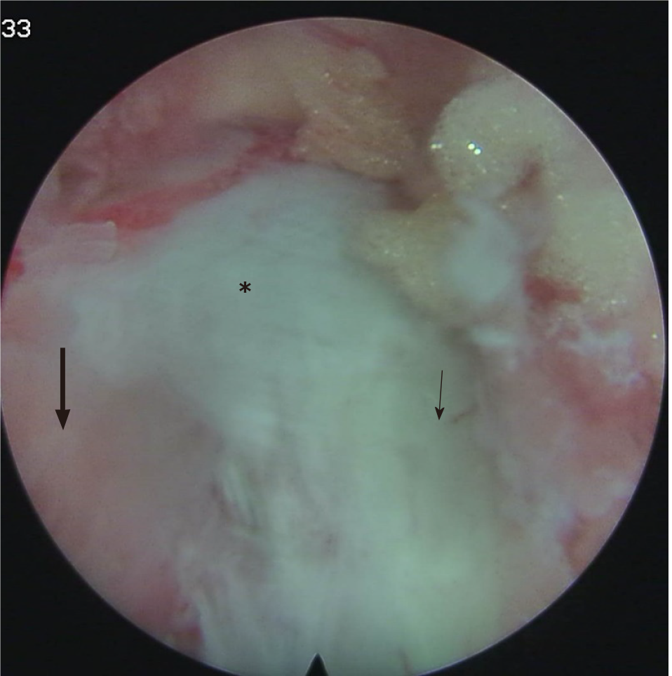
Figure 3 L5 inferior articular process (thin arrow), S1 superior articular process (thick arrow), the joint capsule was exposed (asterisk).
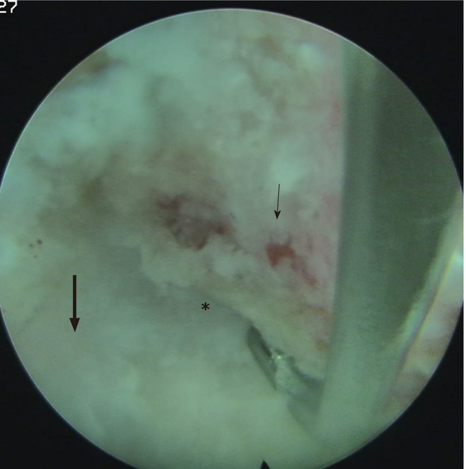
Figure 4 After the operation, the joint space was clear, and no bleeding points existed.
L5 inferior articular process (thin arrow), S1 superior articular process (thick arrow), the joint capsule was exposed (asterisk).
- Citation: Yuan HJ, Wang CY, Wang YF. Endoscopic joint capsule and articular process excision to treat lumbar facet joint syndrome: A case report. World J Clin Cases 2021; 9(28): 8545-8551
- URL: https://www.wjgnet.com/2307-8960/full/v9/i28/8545.htm
- DOI: https://dx.doi.org/10.12998/wjcc.v9.i28.8545


