Copyright
©The Author(s) 2021.
World J Clin Cases. Oct 6, 2021; 9(28): 8498-8503
Published online Oct 6, 2021. doi: 10.12998/wjcc.v9.i28.8498
Published online Oct 6, 2021. doi: 10.12998/wjcc.v9.i28.8498
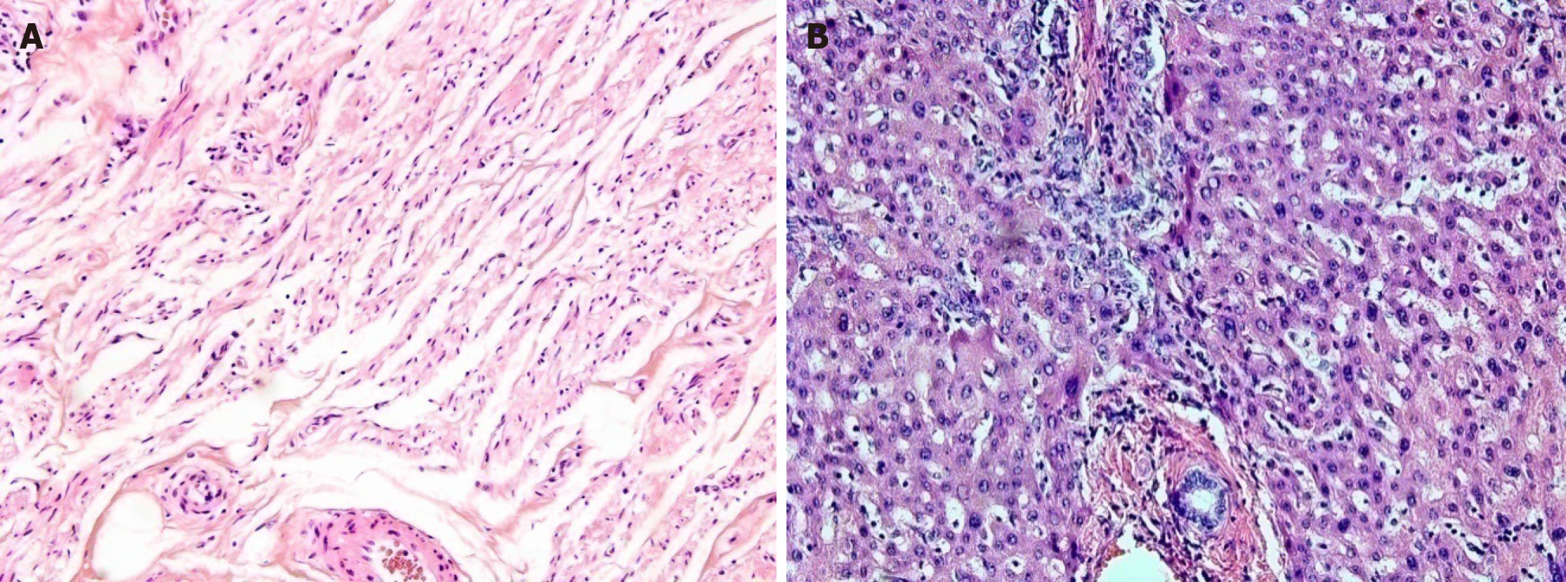
Figure 1 Histology of malignant melanoma.
A: Eyelid tissue; B: Liver tissue.
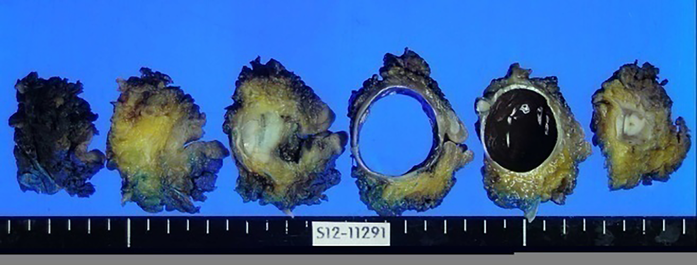
Figure 2 Macroscopic appearance of the uveal melanoma affecting the left eye and eyelid.
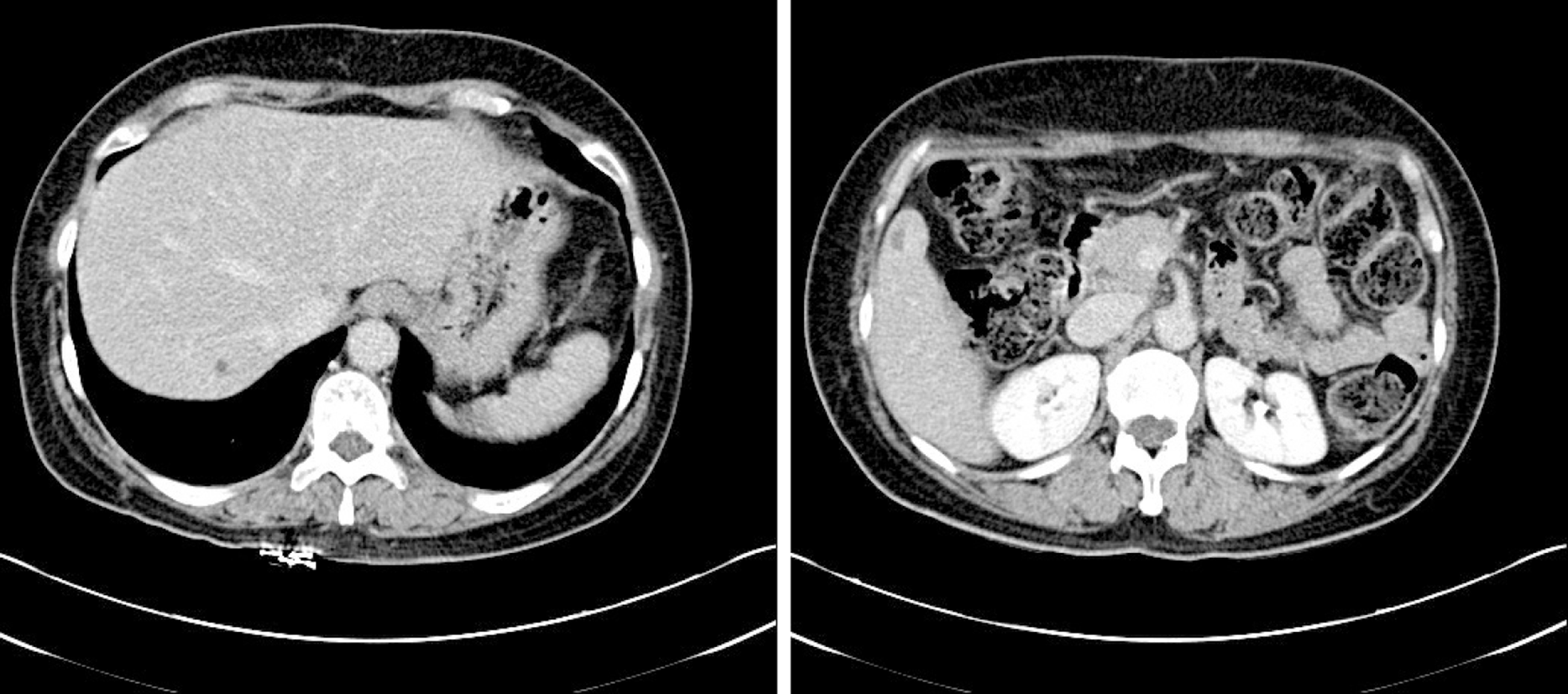
Figure 3 Computed Tomography images of the liver showing multiple liver metastases.
Representative images.
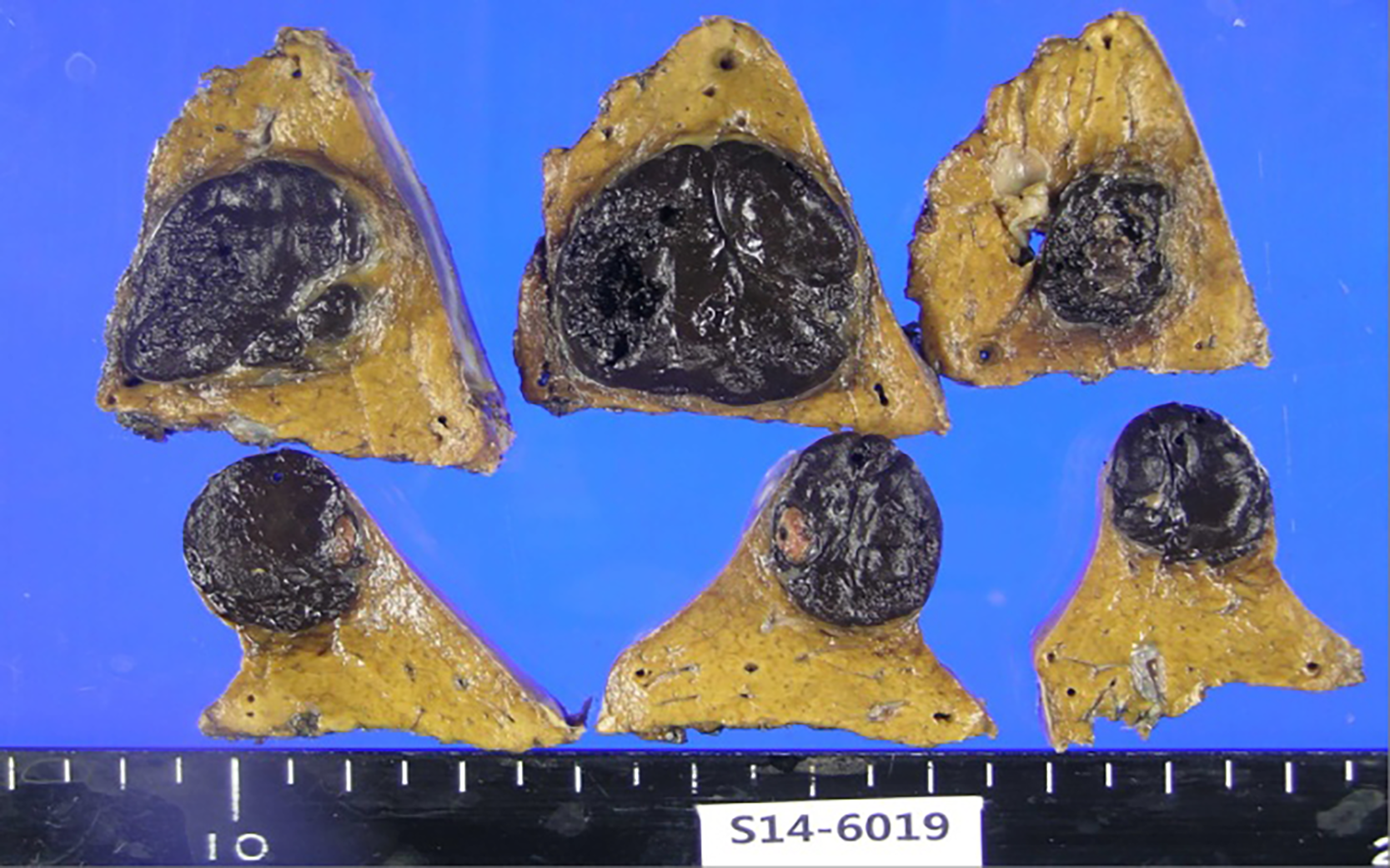
Figure 4 Macroscopic appearance of liver segments with pigmented deposits of metastatic uveal melanoma.
- Citation: Kim YH, Choi NK. Surgical treatment of liver metastasis with uveal melanoma: A case report. World J Clin Cases 2021; 9(28): 8498-8503
- URL: https://www.wjgnet.com/2307-8960/full/v9/i28/8498.htm
- DOI: https://dx.doi.org/10.12998/wjcc.v9.i28.8498


