Copyright
©The Author(s) 2021.
World J Clin Cases. Oct 6, 2021; 9(28): 8470-8475
Published online Oct 6, 2021. doi: 10.12998/wjcc.v9.i28.8470
Published online Oct 6, 2021. doi: 10.12998/wjcc.v9.i28.8470
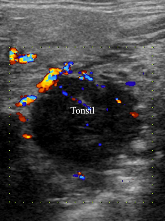
Figure 1 Ultrasound showed a hypoechoic round mass in the right tonsil with well-defined margins, homogeneous echogenicity, and rich irregular blood flow.
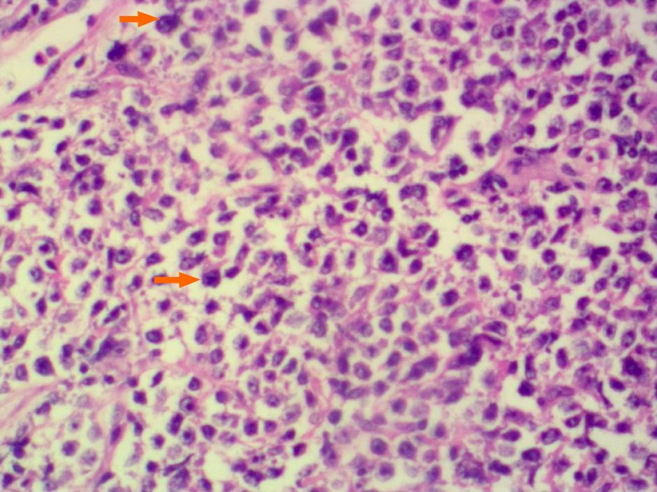
Figure 2 Photomicrograph of a diffuse large B cell lymphoma demonstrating that regional tumor cells (orange arrows) are mononuclear or multinucleated, resembling histiocytes and Reed-Sternberg cells (400 ×, hematoxylin-eosin staining).
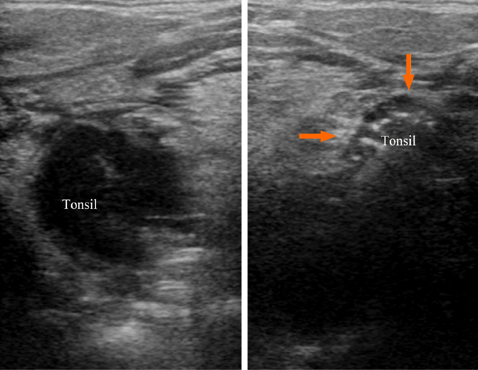
Figure 3 Comparison of bilateral tonsils.
The normal tonsil (indicated by the arrows) presents as homogeneously ovoid echogenic soft tissue with stripes and linear echo inside, while the right tonsil as a hypoechoic round mass with the loss of normal striated pattern.
- Citation: Jiang R, Zhang HM, Wang LY, Pian LP, Cui XW. Ultrasound features of primary non-Hodgkin’s lymphoma of the palatine tonsil: A case report. World J Clin Cases 2021; 9(28): 8470-8475
- URL: https://www.wjgnet.com/2307-8960/full/v9/i28/8470.htm
- DOI: https://dx.doi.org/10.12998/wjcc.v9.i28.8470


