Copyright
©The Author(s) 2021.
World J Clin Cases. Sep 6, 2021; 9(25): 7605-7613
Published online Sep 6, 2021. doi: 10.12998/wjcc.v9.i25.7605
Published online Sep 6, 2021. doi: 10.12998/wjcc.v9.i25.7605
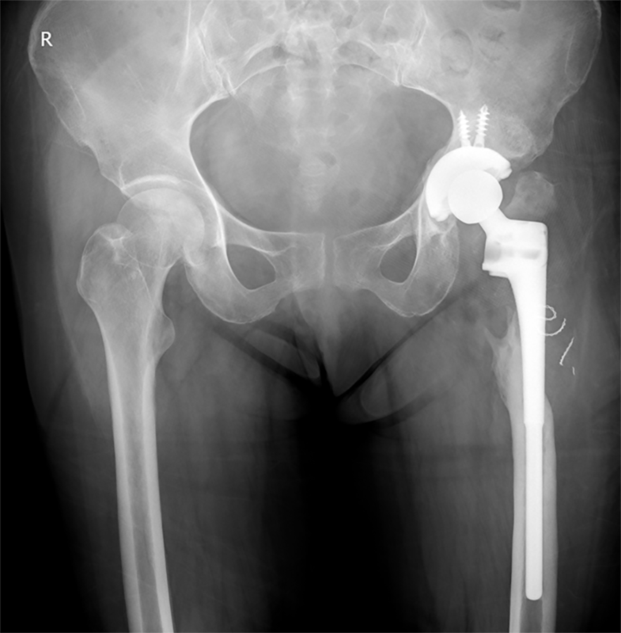
Figure 1 X-ray film, anteroposterior plane.
The left artificial hip had a severe proximal femoral defect.
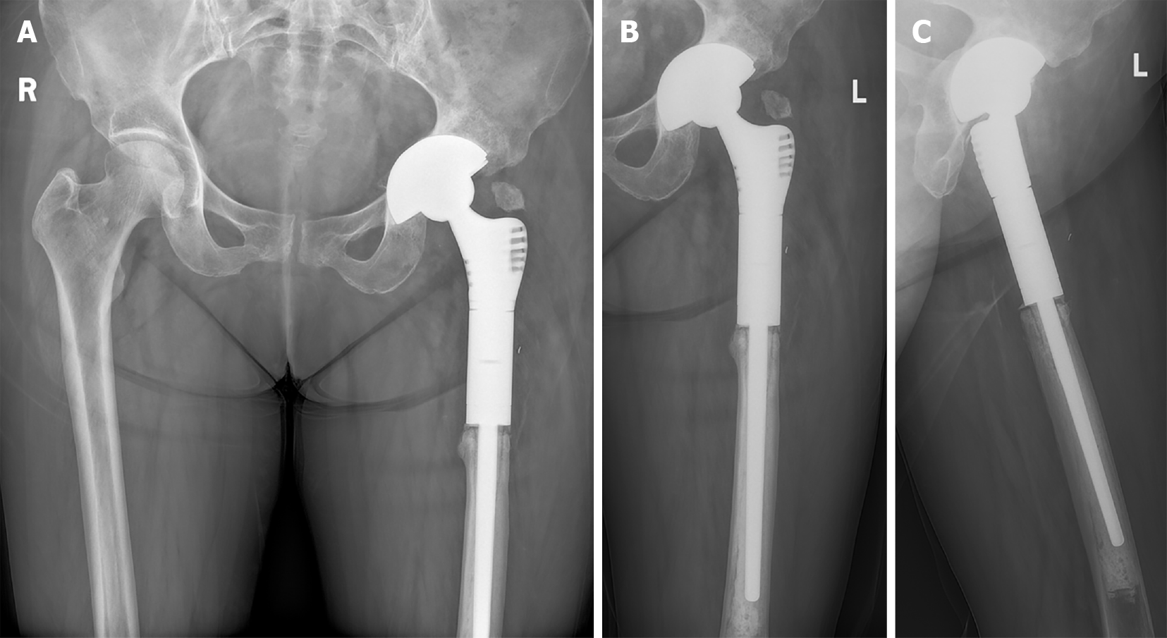
Figure 2 Radiographs of the pelvic taken 20 mo after the second-stage surgery.
A: Pelvic plane; B: Anteroposterior plane; C: Lateral plane.
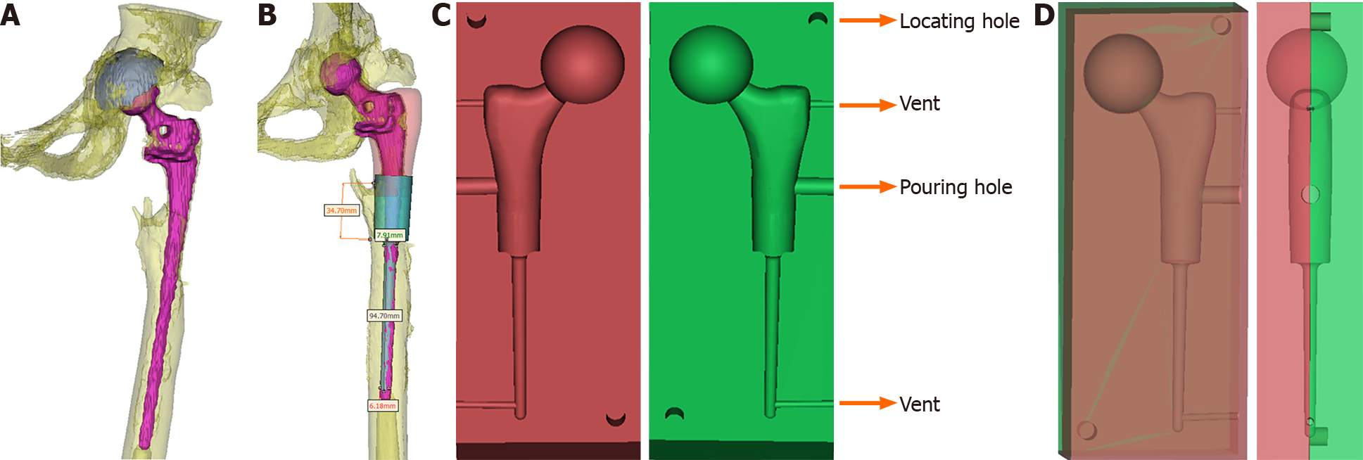
Figure 3 Design process of the customized spacer molds.
A: Reconstruction image of the prosthesis and left hip; B: The blueprint of the spacer according to the reconstruction image; C: The blueprint of the customized spacer molds; D: The blueprint of the assembled spacer molds.
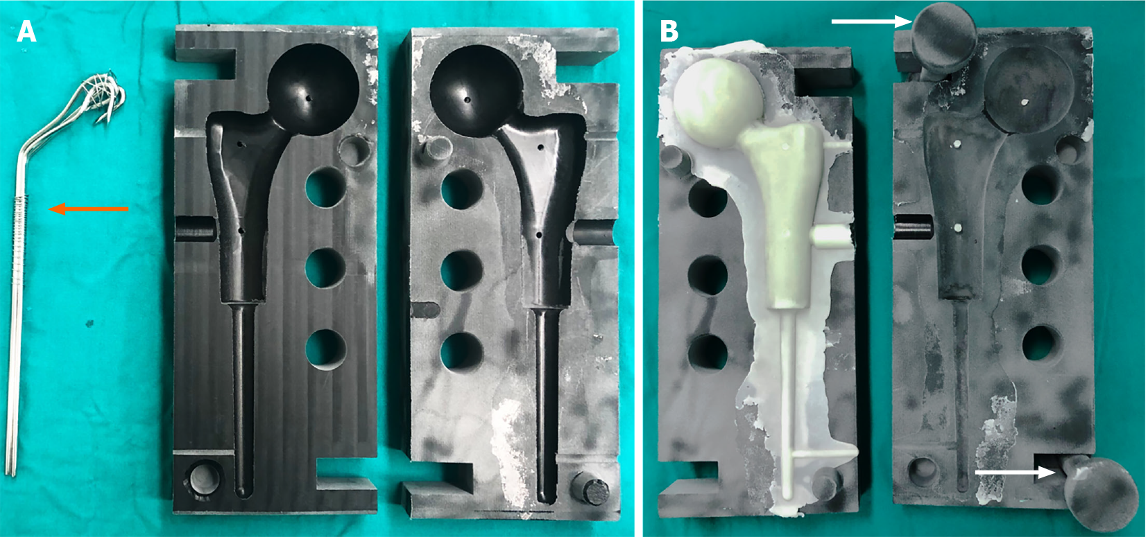
Figure 4 Molding process of the custom-made spacer cement.
A: The molds and the binding bundle of Kirschner wires (orange arrow); B: Spacer cement and the pressure rods (white arrow).
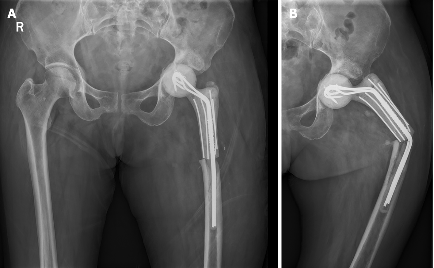
Figure 5 Radiographs of the pelvic taken the day after the first-stage revision (anteroposterior plane) (A) and the left hip taken 6 mo after the first-stage revision (B).
The spacer stem was bent (anteroposterior plane).
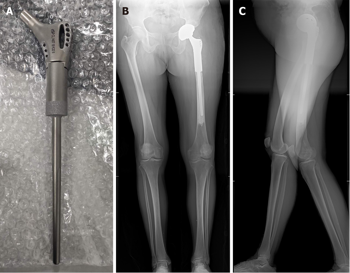
Figure 6 The custom-made prosthesis with porous design, and full lower limb radiographs taken the day after the second-stage revision.
A: The custom-made prosthesis with porous design; B: Anteroposterior plane; C: Lateral plane.
- Citation: Liu YB, Pan H, Chen L, Ye HN, Wu CC, Wu P, Chen L. Total hip revision with custom-made spacer and prosthesis: A case report. World J Clin Cases 2021; 9(25): 7605-7613
- URL: https://www.wjgnet.com/2307-8960/full/v9/i25/7605.htm
- DOI: https://dx.doi.org/10.12998/wjcc.v9.i25.7605


