Copyright
©The Author(s) 2021.
World J Clin Cases. Sep 6, 2021; 9(25): 7600-7604
Published online Sep 6, 2021. doi: 10.12998/wjcc.v9.i25.7600
Published online Sep 6, 2021. doi: 10.12998/wjcc.v9.i25.7600
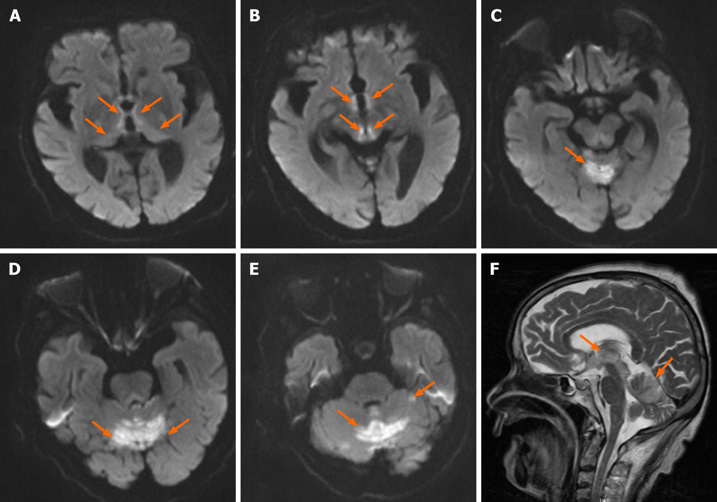
Figure 1 Diffusion-weighted imaging.
A-E: Axial diffusion-weighted imaging showing bilateral and symmetric high signals at the level of the periventricular regions of the third ventricle, medial portion of bilateral thalamus, corpora quadrigemina (A and B), vermis, (C) and cerebellar hemispheres (D and E); F: Sagittal T2-weighted imaging lesions.
- Citation: Nie T, He JL. Wernicke's encephalopathy in a rectal cancer patient with atypical radiological features: A case report. World J Clin Cases 2021; 9(25): 7600-7604
- URL: https://www.wjgnet.com/2307-8960/full/v9/i25/7600.htm
- DOI: https://dx.doi.org/10.12998/wjcc.v9.i25.7600


