Copyright
©The Author(s) 2021.
World J Clin Cases. Sep 6, 2021; 9(25): 7564-7571
Published online Sep 6, 2021. doi: 10.12998/wjcc.v9.i25.7564
Published online Sep 6, 2021. doi: 10.12998/wjcc.v9.i25.7564
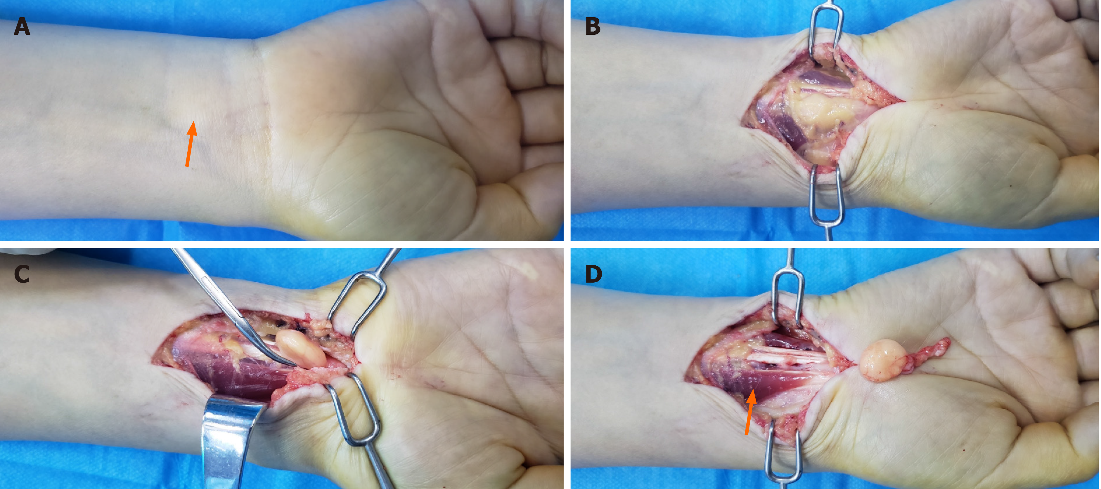
Figure 1 Operating images of the entire surgical process.
A: Preoperatively, there was an intumescence at the distal end of the forearm (orange arrow); B and C: The mass was revealed layer-by-layer during the operation; D: The mass and surrounding fascia were completely removed, and the muscle belly of the flexor digitorum superficialis was abnormally shifted down (orange arrow).
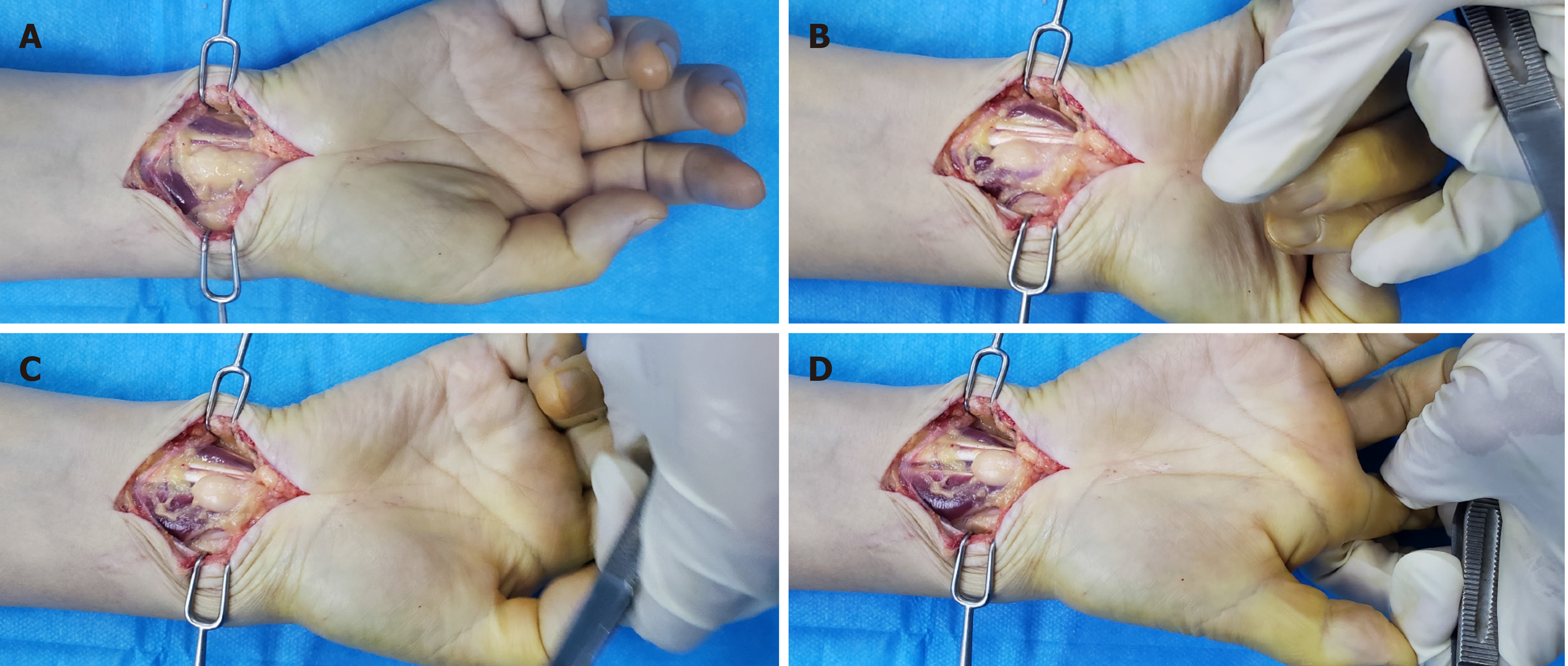
Figure 2 Entrapment symptoms appeared within the carpal tunnel during finger flexion and extension.
A: The tumor involved the flexor digitorum superficialis of the index finger in the carpal tunnel; B: In the passive flexion position of the index finger, the mass moved proximally with the flexor digitorum superficialis; C and D: In the extension position of the index finger, the mass moved with the flexor digitorum superficialis and extended into the carpal tunnel. In the overextension position, the mass further moved into the carpal tunnel, extruding the common flexor tendon sheath and the median nerve.
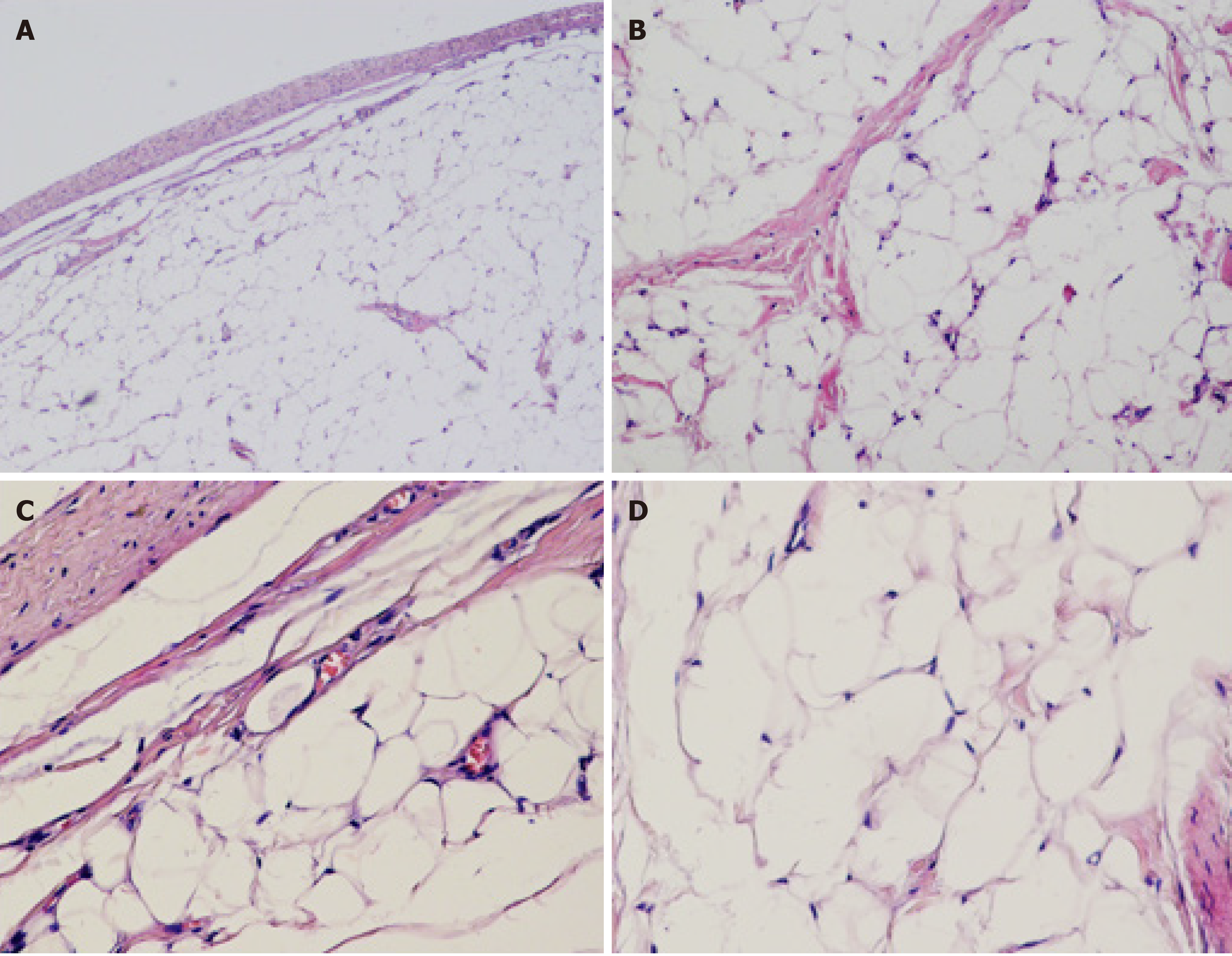
Figure 3 Microscopic observations revealed mature adipocytes and some muscle fiber tissues, with no adipoblasts and abnormal mitotic cells in the tumor.
A: Under low magnification (10 ×); B: Under magnification (100 ×); C and D: Under high magnification (200 ×).
- Citation: Huang C, Jin HJ, Song DB, Zhu Z, Tian H, Li ZH, Qu WR, Li R. Trigger finger at the wrist caused by an intramuscular lipoma within the carpal tunnel: A case report. World J Clin Cases 2021; 9(25): 7564-7571
- URL: https://www.wjgnet.com/2307-8960/full/v9/i25/7564.htm
- DOI: https://dx.doi.org/10.12998/wjcc.v9.i25.7564


