Copyright
©The Author(s) 2021.
World J Clin Cases. Sep 6, 2021; 9(25): 7558-7563
Published online Sep 6, 2021. doi: 10.12998/wjcc.v9.i25.7558
Published online Sep 6, 2021. doi: 10.12998/wjcc.v9.i25.7558
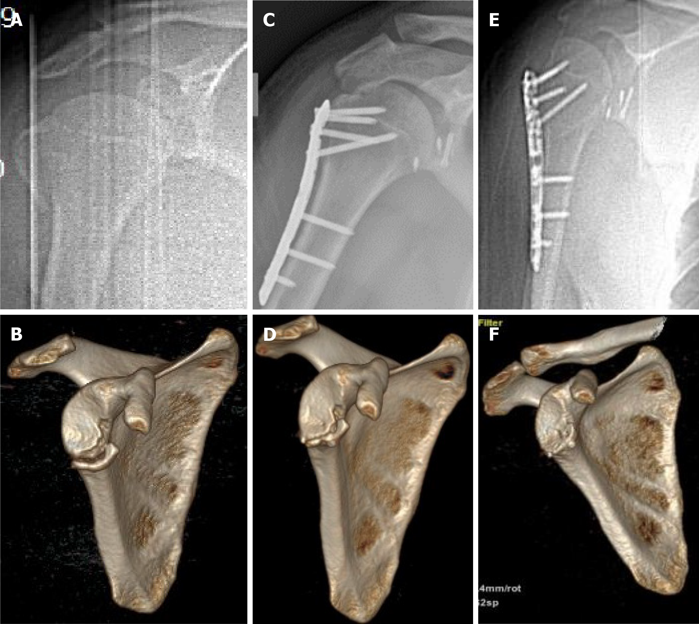
Figure 1 Preoperative and postoperative radiography images of the patient.
A: Preoperative X-ray; B: Preoperative three-dimensional (3D) reconstruction; C: X-ray image at 1 wk postoperatively; D: 3D reconstruction at 1 wk postoperatively; E: X-ray image at 12 mo postoperatively; F: 3D reconstruction at 12 mo postoperatively.
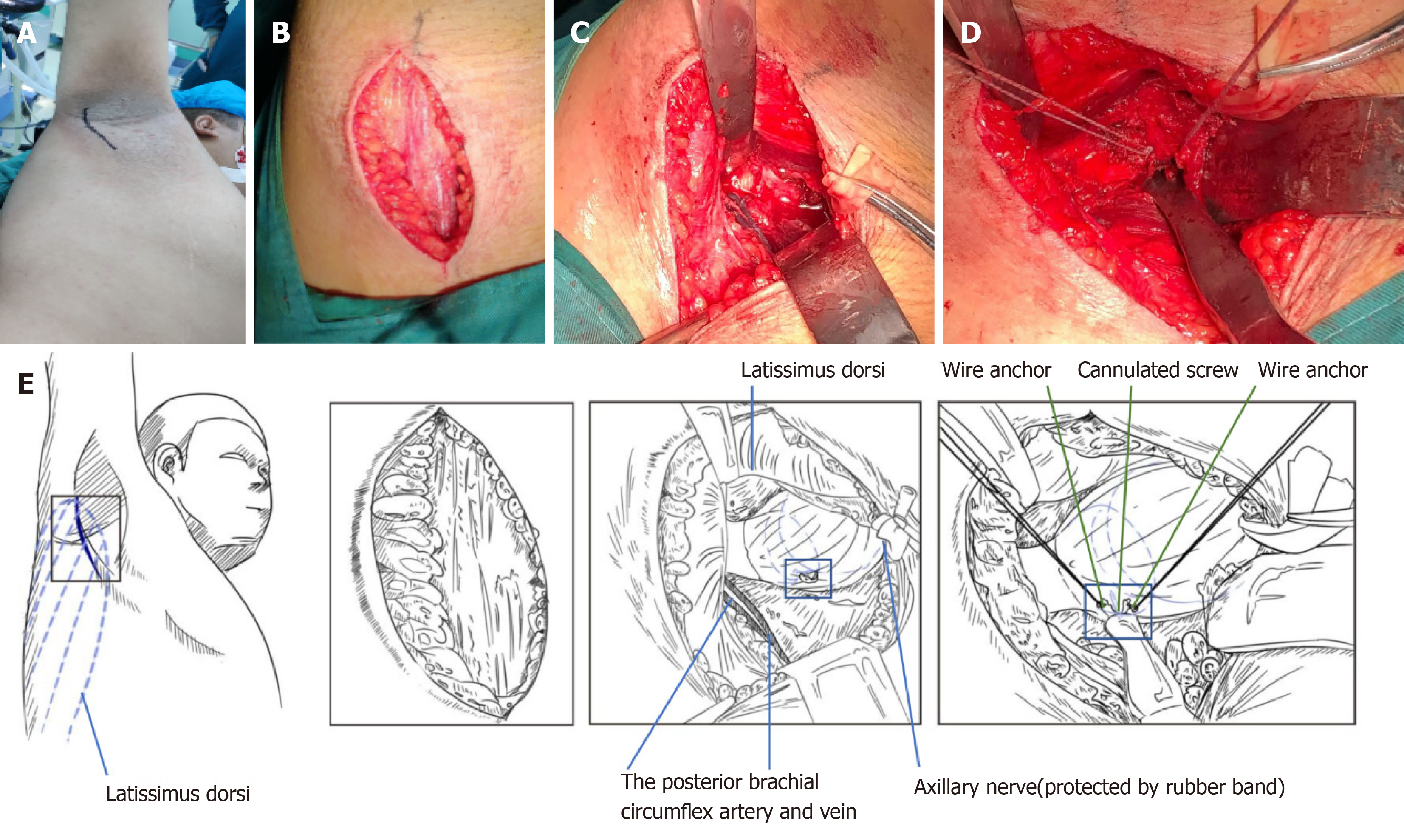
Figure 2 Intraoperative photos and sketches.
A: Surgical marker; B: A longitudinal incision was made in the armpit, followed by exposure of the anterior edge of the latissimus dorsi by separating subcutaneous tissues; C: The posterior brachial circumflex artery and vein, under the axillary nerve, were exposed. The blood vessels and nerves were protected by a tender traction; D: The fracture block was fixed with one cannulated screw and two wire anchors were used to strengthen the fixation; E: The sketches more vividly describes the whole operation process.
- Citation: Jia X, Zhou FL, Zhu YH, Jin DJ, Liu WX, Yang ZC, Liu RP. Treatment of lower part of glenoid fractures through a novel axillary approach: A case report. World J Clin Cases 2021; 9(25): 7558-7563
- URL: https://www.wjgnet.com/2307-8960/full/v9/i25/7558.htm
- DOI: https://dx.doi.org/10.12998/wjcc.v9.i25.7558


