Copyright
©The Author(s) 2021.
World J Clin Cases. Sep 6, 2021; 9(25): 7542-7550
Published online Sep 6, 2021. doi: 10.12998/wjcc.v9.i25.7542
Published online Sep 6, 2021. doi: 10.12998/wjcc.v9.i25.7542
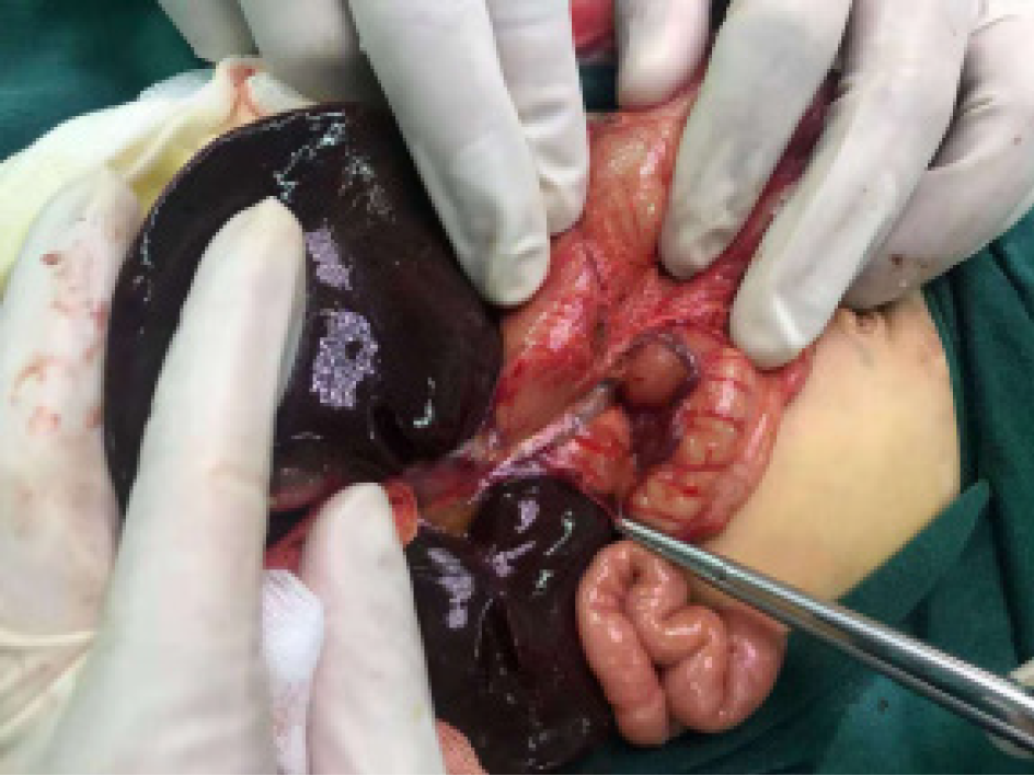
Figure 1 The child underwent laparoscopic exploration under general anesthesia.
During the operation, the portal vein was located at the anterior edge of the duodenum.
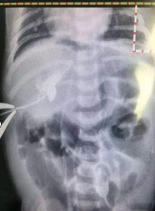
Figure 2 Intraoperative cholangiography showed that the intrahepatic bile duct was visualized by percutaneous puncture catheter-based injection of the contrast agent, but the biliary tract system was not clearly visualized, the duodenum was not visualized, and there was no contrast agent in the abdominal cavity.
- Citation: Xiang XL, Cai P, Zhao JG, Zhao HW, Jiang YL, Zhu ML, Wang Q, Zhang RY, Zhu ZW, Chen JL, Gu ZC, Zhu J. Neonatal biliary atresia combined with preduodenal portal vein: A case report. World J Clin Cases 2021; 9(25): 7542-7550
- URL: https://www.wjgnet.com/2307-8960/full/v9/i25/7542.htm
- DOI: https://dx.doi.org/10.12998/wjcc.v9.i25.7542


