Copyright
©The Author(s) 2021.
World J Clin Cases. Sep 6, 2021; 9(25): 7520-7526
Published online Sep 6, 2021. doi: 10.12998/wjcc.v9.i25.7520
Published online Sep 6, 2021. doi: 10.12998/wjcc.v9.i25.7520
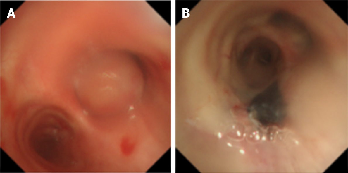
Figure 1 Bronchoscopic images of the main carina.
A: Tumorous endobronchial lesion located in the right main bronchus; B: After removal of the right main mass.
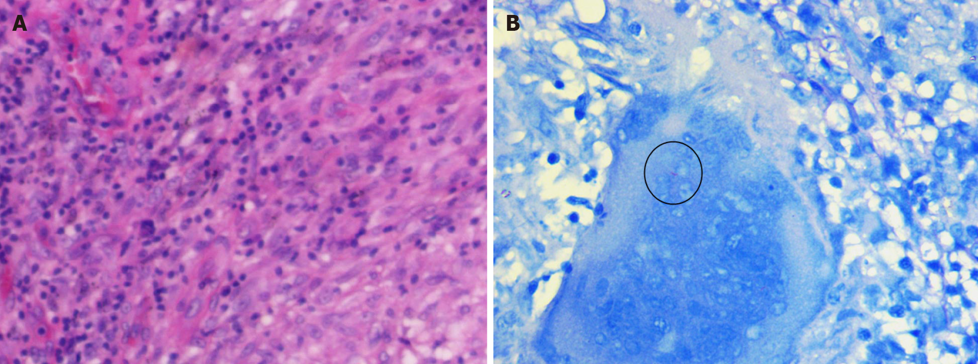
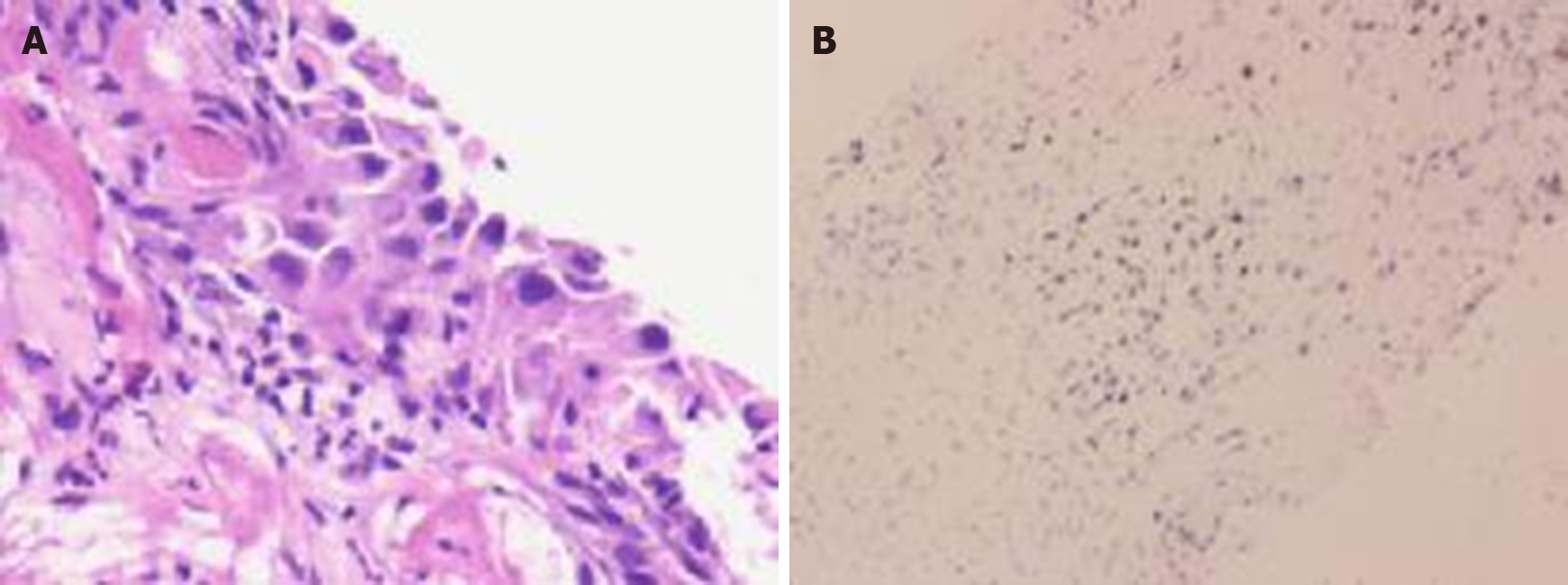
Figure 2 Bronchial biopsy.
A: Granulomatous inflammation with caseification, which is consistent with tuberculosis (Hematoxylin and eosin stain, × 200); B: Biopsy staining showed positivity for acid-fast bacilli (Ziehl-Nielsen staining, × 1000).
Figure 3 Imaging changes of the mass before treatment.
A: Reconstructed thoracic computed tomography (CT) image; B: Enhanced chest CT in mediastinal window; C: Axial CT in lung window showing narrowing of the main bronchus and airspace consolidation in the right bronchus, without parenchymal infiltration or pneumonia.
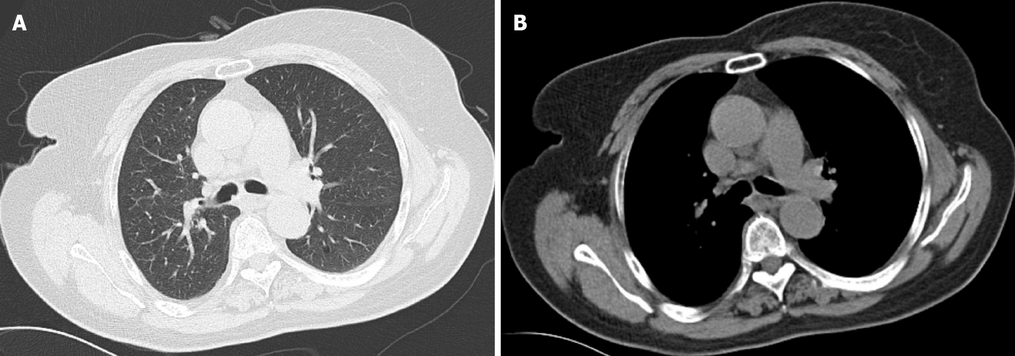
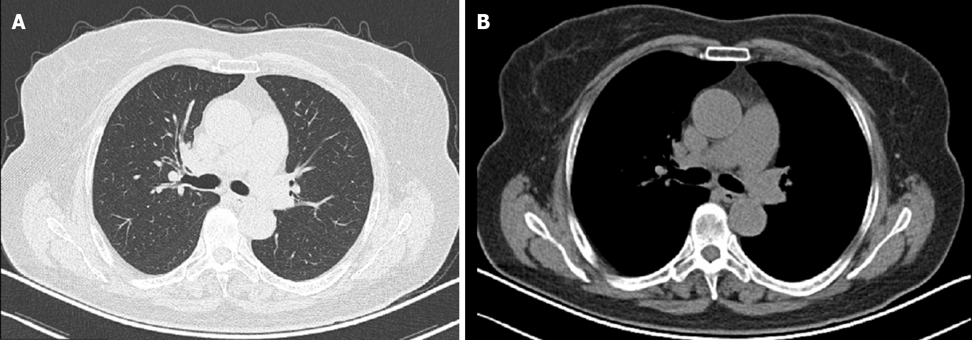
Figure 4 Chest computed tomography images (December 23, 2019) showing a 5 mm × 4 mm lung mass in the right main bronchus.
A: Lung window; B: Mediastinal window.
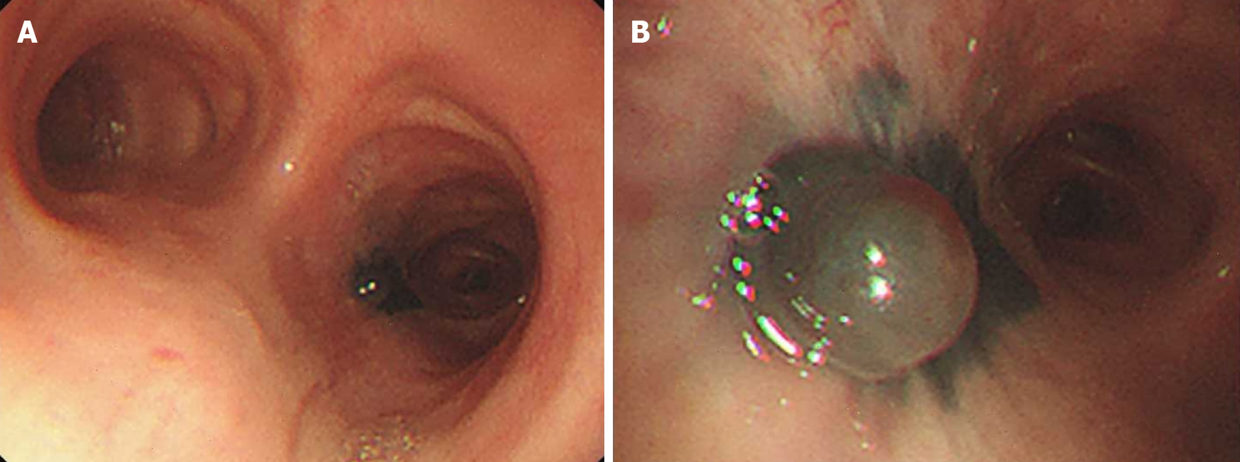
Figure 5 Bronchoscopy (December 23, 2019).
A and B: A black mucosa was found in the right main bronchus.
Figure 6 Histological and immunohistochemical examinations.
A: The right main bronchus mass was squamous cell carcinoma (hematoxylin and eosin staining, × 200); B: The tumor cells were positive for P63, P40, CK7, CK(Pan), Ki67, and p53 but negative for TTF-1, NapsinA, SOX2, SALL4, and CD34 (× 200).
Figure 7 Chest computed tomography images (March 20, 2020) showing complete resolution of the lesion in the right main bronchus.
A: Lung window; B: Mediastinal window.
- Citation: Jiang H, Li YQ. Coexistence of tuberculosis and squamous cell carcinoma in the right main bronchus: A case report. World J Clin Cases 2021; 9(25): 7520-7526
- URL: https://www.wjgnet.com/2307-8960/full/v9/i25/7520.htm
- DOI: https://dx.doi.org/10.12998/wjcc.v9.i25.7520


