Copyright
©The Author(s) 2021.
World J Clin Cases. Sep 6, 2021; 9(25): 7504-7511
Published online Sep 6, 2021. doi: 10.12998/wjcc.v9.i25.7504
Published online Sep 6, 2021. doi: 10.12998/wjcc.v9.i25.7504
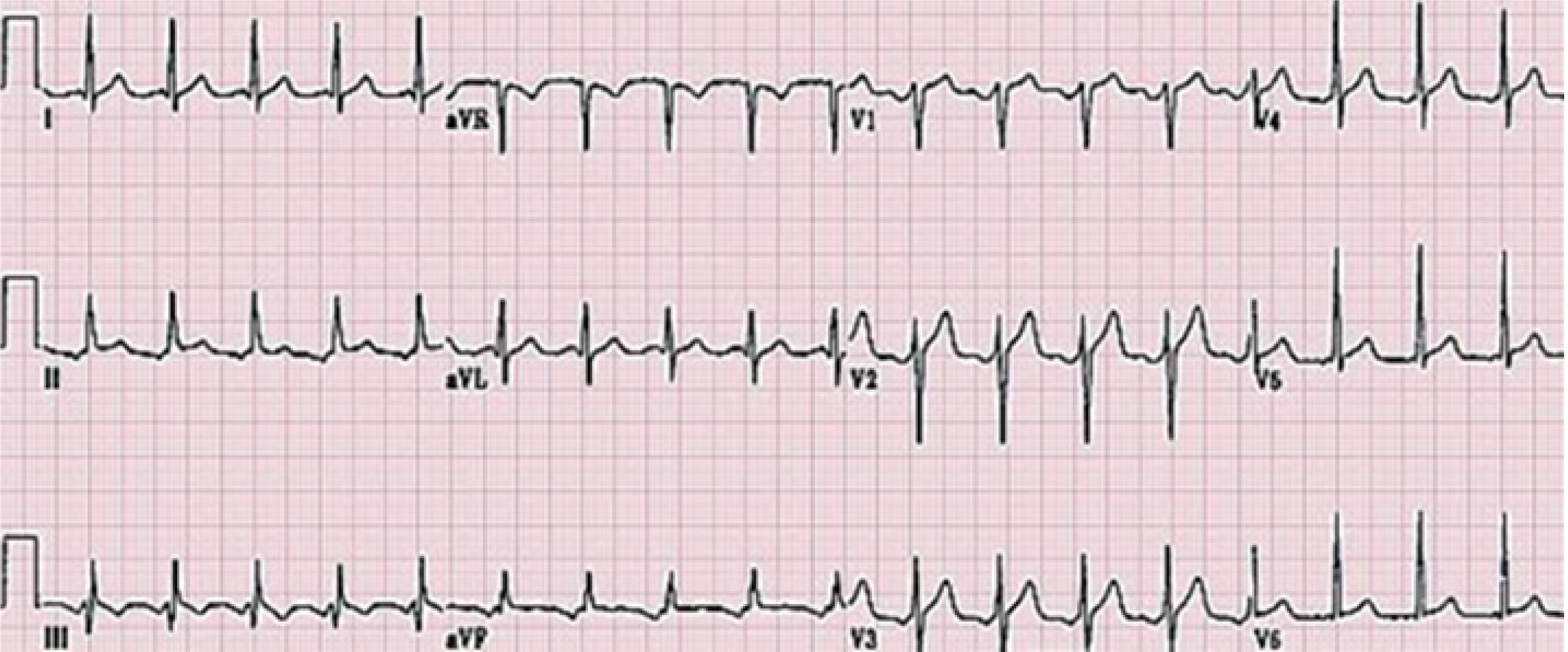
Figure 1 Electrocardiograph showing sinus tachycardia.
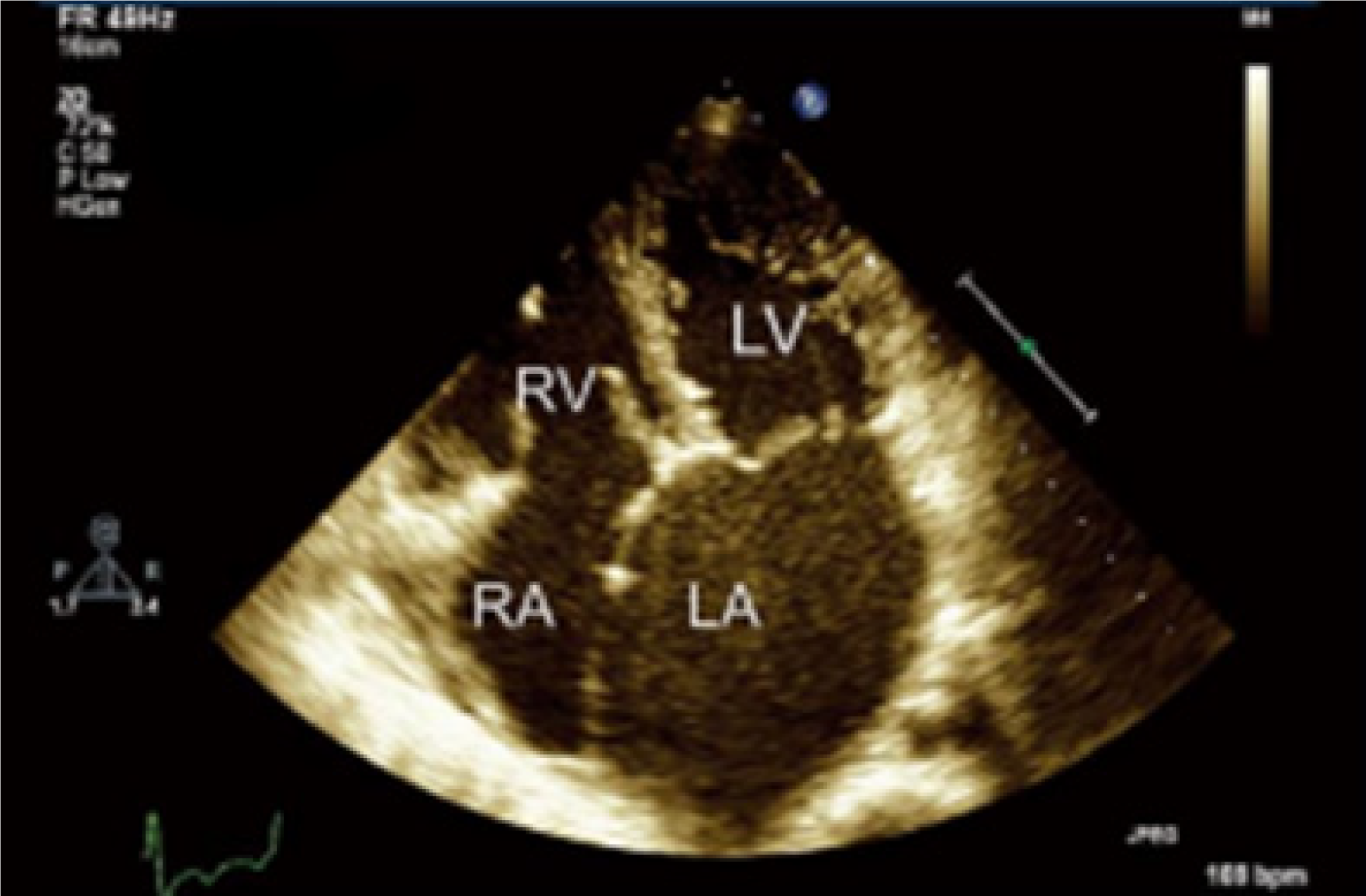
Figure 2 Echocardiography showed diffuse hypokinesis of the left ventricle with a 38% ejection fraction.
LV: Left ventricle; RV: Right ventricle; RA: Right atrium; LA: Left atrium.
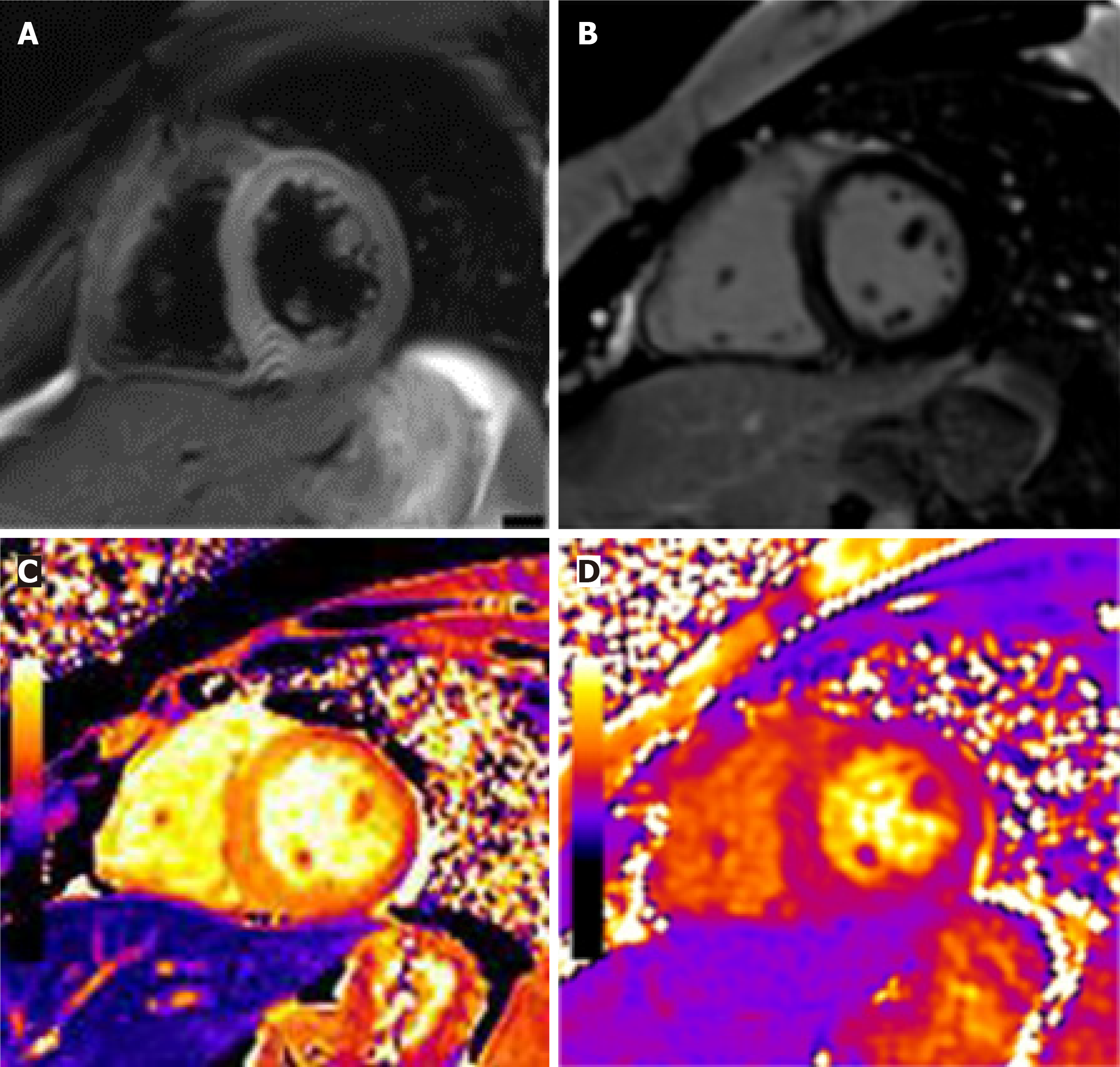
Figure 3 T2-weighted images, late gadolinium enhancement, and T1 and T2 mapping in this patient.
Mapping of the native T1 values of the left ventricle showed a diffusely enhanced T1 value of approximately 1380 msec. T2 = 45 ms. A: Edema ratio; B: Late gadolinium enhancement; C: T1; D: T2.
- Citation: Fu LY, Zhang HB. Effective treatment of polyneuropathy, organomegaly, endocrinopathy, M-protein, and skin changes syndrome with congestive heart failure: A case report. World J Clin Cases 2021; 9(25): 7504-7511
- URL: https://www.wjgnet.com/2307-8960/full/v9/i25/7504.htm
- DOI: https://dx.doi.org/10.12998/wjcc.v9.i25.7504


