Copyright
©The Author(s) 2021.
World J Clin Cases. Sep 6, 2021; 9(25): 7490-7497
Published online Sep 6, 2021. doi: 10.12998/wjcc.v9.i25.7490
Published online Sep 6, 2021. doi: 10.12998/wjcc.v9.i25.7490
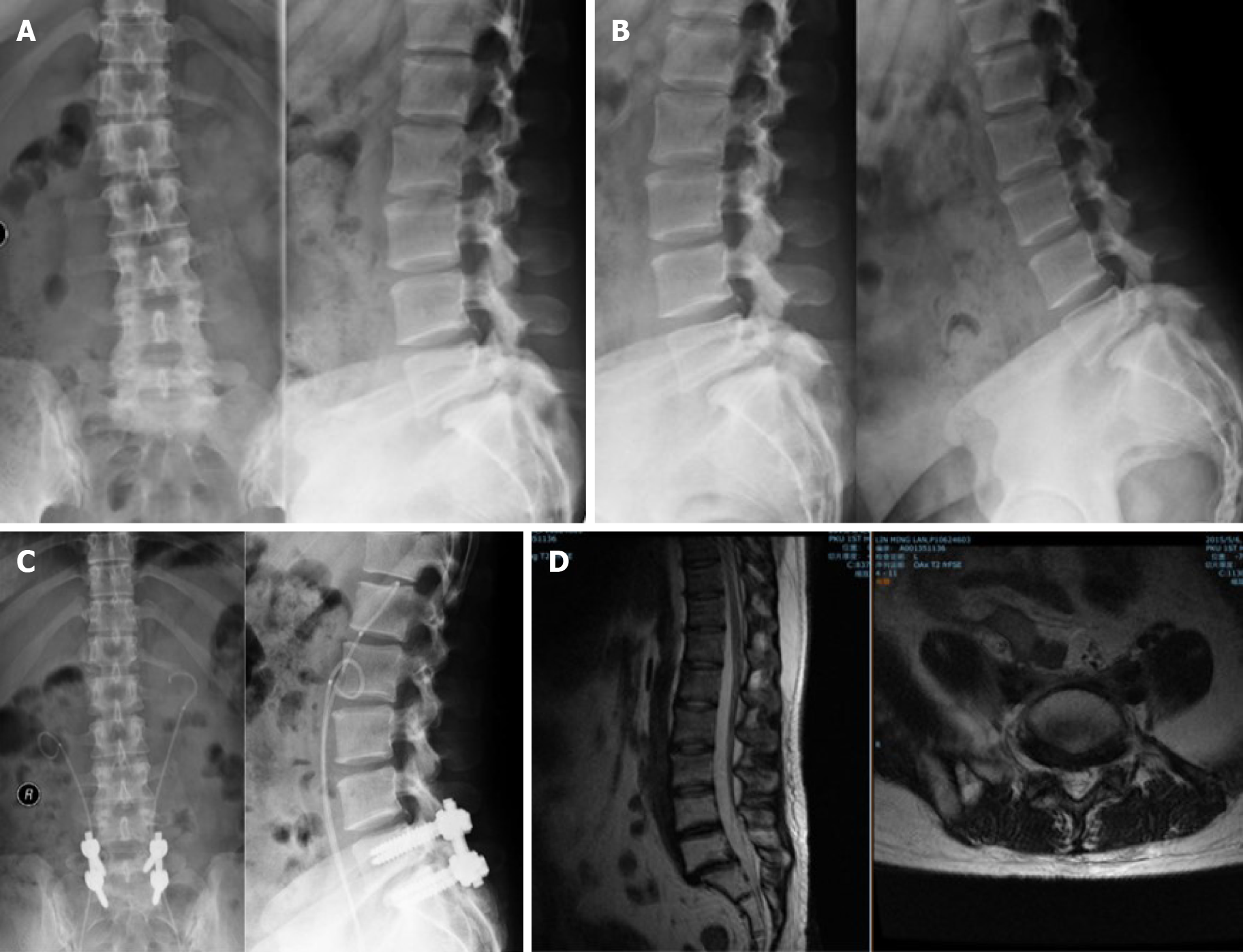
Figure 1 Peri-operative radiography.
A: Preoperative anteroposterior and lateral radiographs; B: Preoperative flexion-extension radiographs; C: Postoperative anteroposterior and lateral radiographs; D: Preoperative magnetic resonance imaging on sagittal and L5/S1 axial planes.
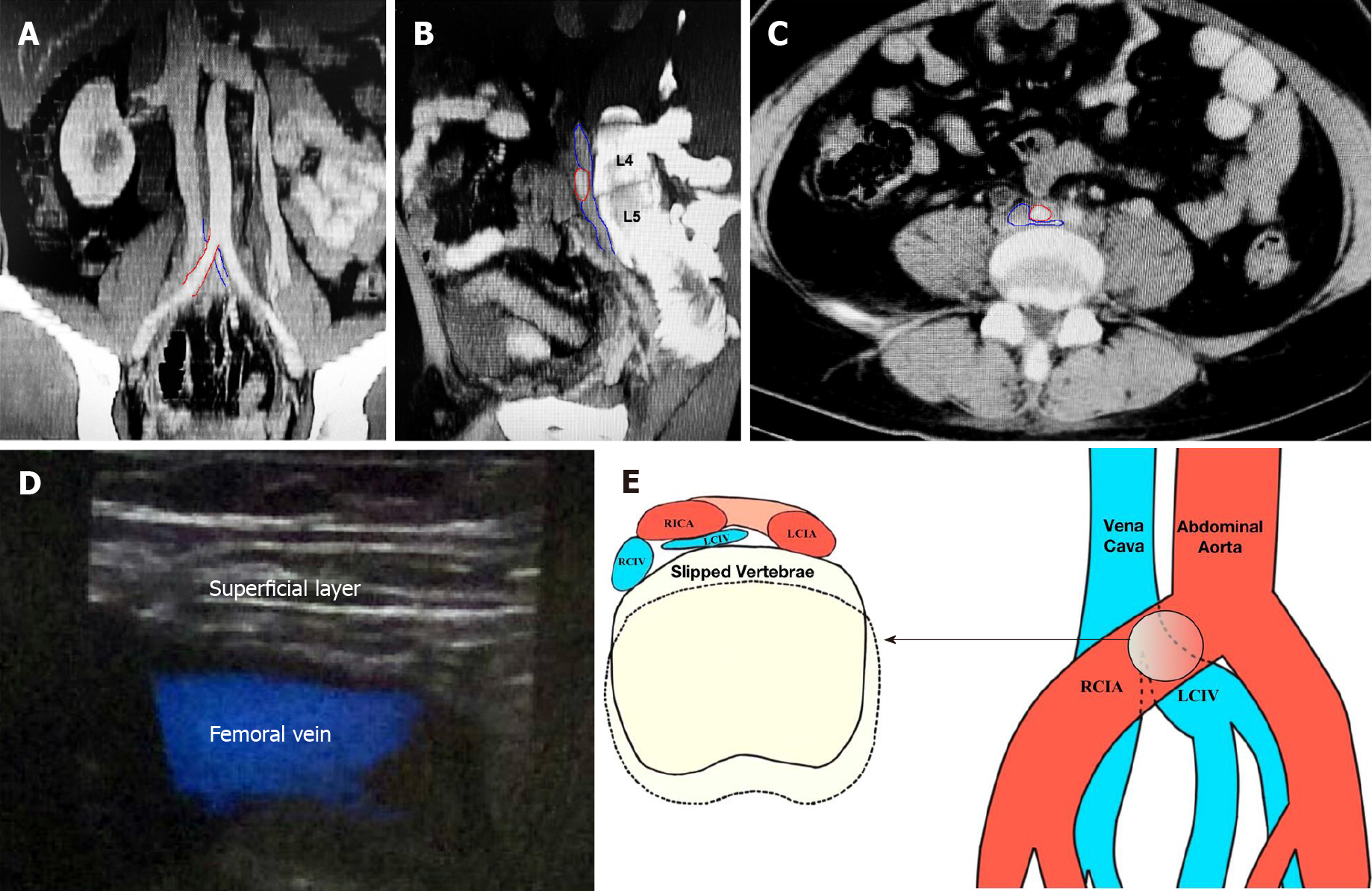
Figure 2 Preoperative May-Thurner syndrome radiography and mechanisms.
A: Coronal plane of computed tomography (CT) showing the right common iliac artery (RCIA, red outline), and left common iliac vein (LCIV, blue outline); B: Adjusted plane of CT shows the RCIA (red outline) and the vena cava and LCIV (blue outline). The RCIA compresses the adjoining part of the vena cava and LCIV against the vertebrae; C: Axial plane of CT showing the RCIA (red outline) and the LCIV (blue outline); D: Preoperative ultrasonography shows dilation of femoral vein without thrombus; E: Mechanisms of May-Thurner syndrome. LCIA: Left common iliac artery; RCIV: Right common iliac vein.
- Citation: Yue L, Fu HY, Sun HL. Acute deep venous thrombosis induced by May-Thurner syndrome after spondylolisthesis surgery: A case report and review of literature. World J Clin Cases 2021; 9(25): 7490-7497
- URL: https://www.wjgnet.com/2307-8960/full/v9/i25/7490.htm
- DOI: https://dx.doi.org/10.12998/wjcc.v9.i25.7490


