Copyright
©The Author(s) 2021.
World J Clin Cases. Sep 6, 2021; 9(25): 7459-7467
Published online Sep 6, 2021. doi: 10.12998/wjcc.v9.i25.7459
Published online Sep 6, 2021. doi: 10.12998/wjcc.v9.i25.7459
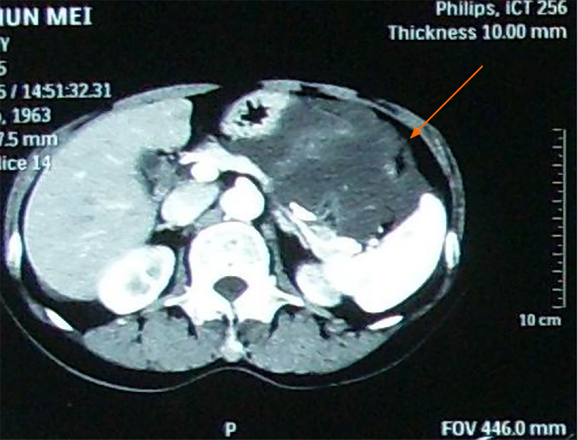
Figure 1 Abdominal contrast-enhanced computed tomography revealed a low density mass in the upper abdomen proximal to the spleen (arrow).
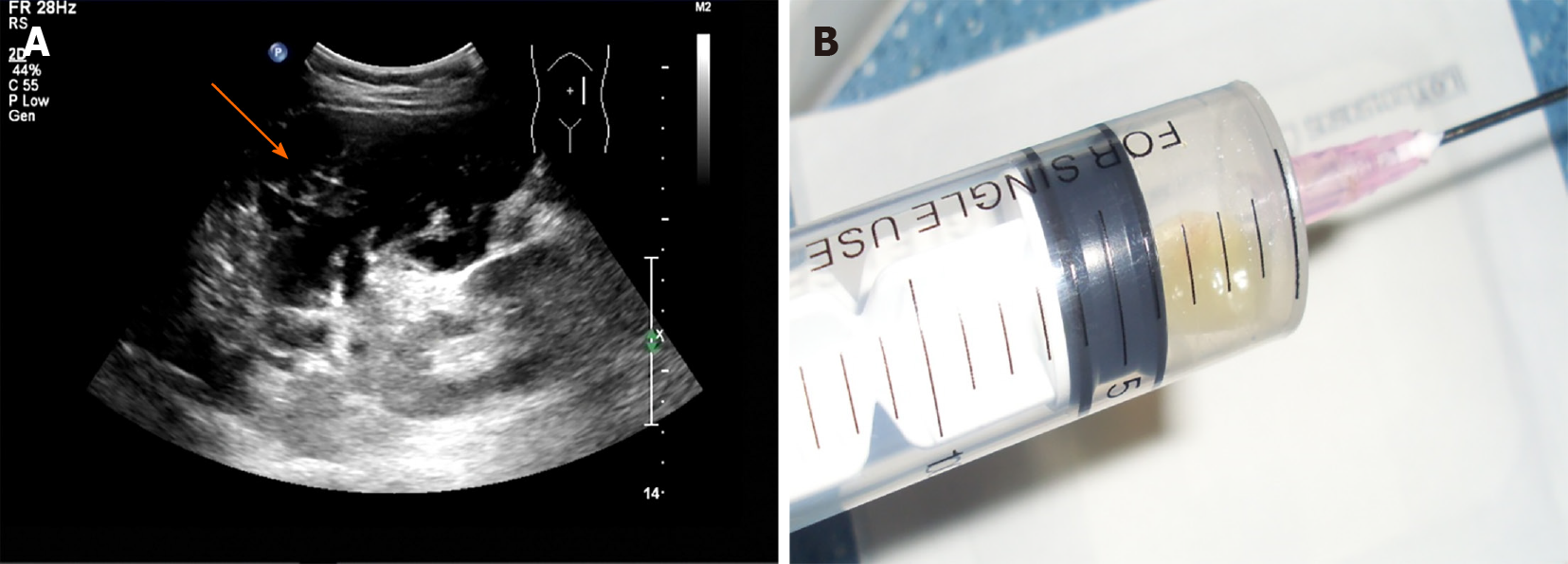
Figure 2 Ultrasound image and transabdominal ultrasound-guided percutaneous aspiration of the mass in the left upper abdomen.
A: A large mass with flocculent and stripe-like echoes (arrow) was detected in the left middle and upper abdomen by ultrasound; B: Yellow gelatinous material was aspirated from the abdomen via transabdominal ultrasound-guided percutaneous aspiration.
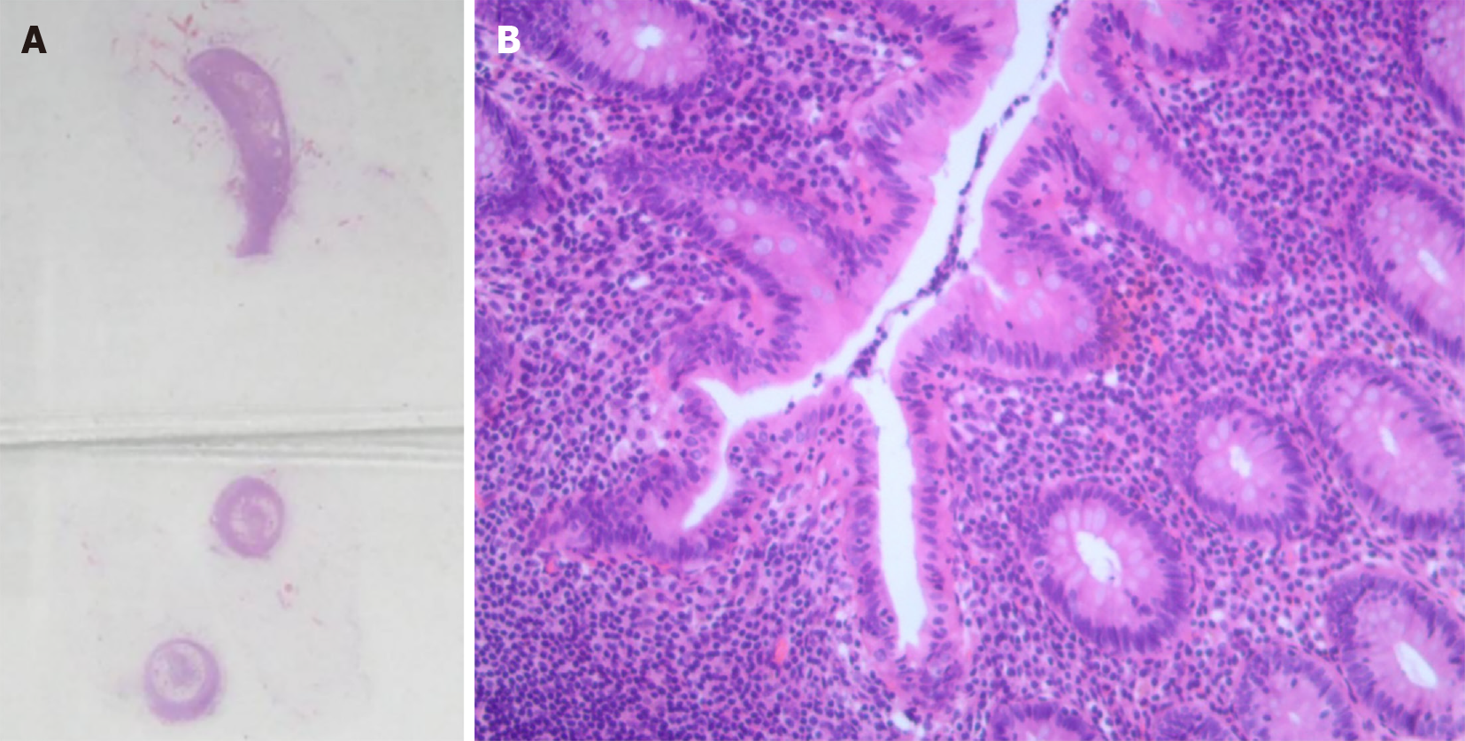
Figure 3 The obtained appendix specimen and its hematoxylin-eosin staining results.
A: Gross pathology of appendix showed a length of 5.0 cm and width of 0.3-0.6 cm in diameter; B: Hematoxylin-eosin staining results of the specimen demonstrated appendicitis obliterans.
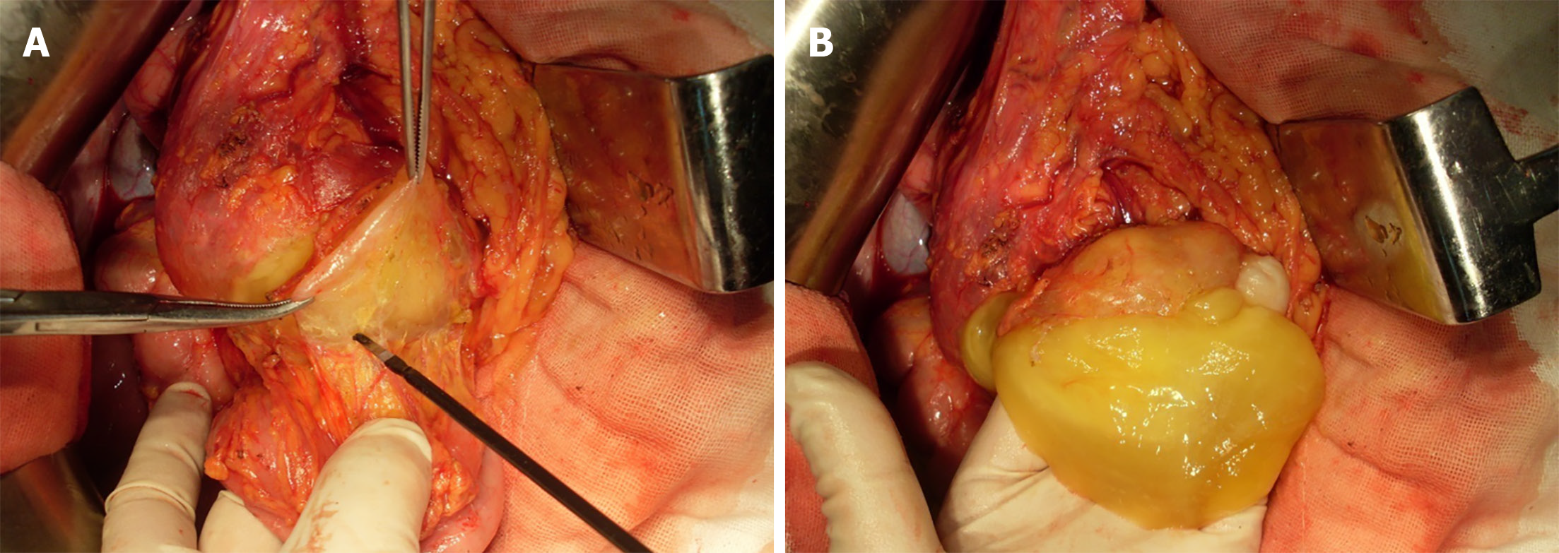
Figure 4 Intraoperative pictures.
A: Characteristic cystic mass (arrow) presented in the anterior lobe of the transverse mesocolon in the left part of the splenic flexure; B: A yellow jelly-like mass existed inside.
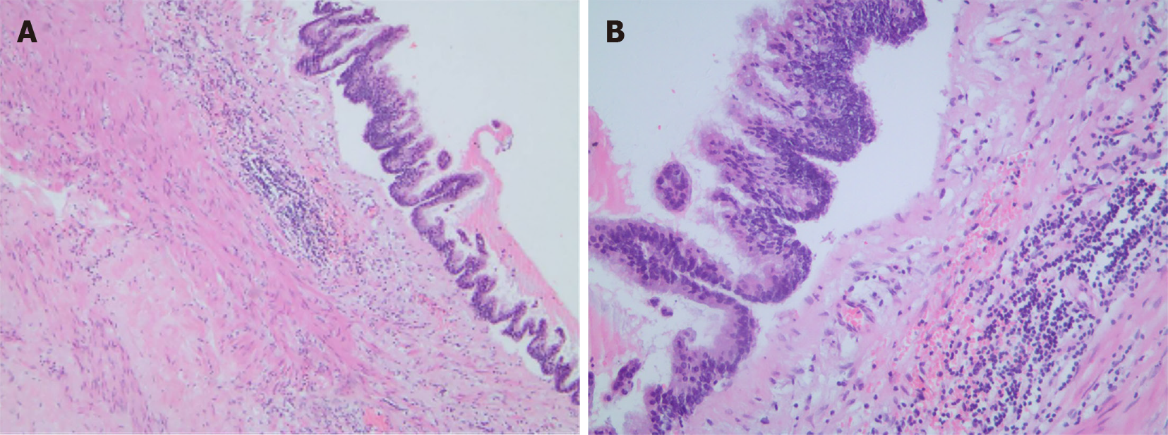
Figure 5 Hematoxylin-eosin staining of the specimen found in the splenic flexure of the colon revealed a cystic mass emanating from the intestinal duplication, with low-grade mucinous epithelial cells lining in the capsule wall.
A: × 40; B: × 200.
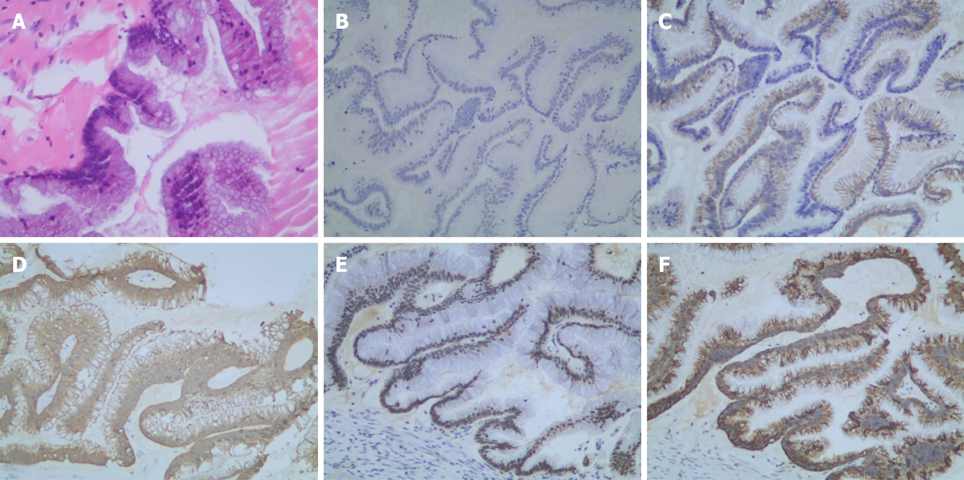
Figure 6 Histologic presentation of low-grade mucinous neoplasm.
A: Hematoxylin-eosin staining of the primary tumor; B-F: Immunohistochemical staining found that the primary tumor was CK-7(–) (B), CK-20(+) (C), Villin(+) (D), CDX-2(+) (E), and MUC-2(+) (F).
- Citation: Han XD, Zhou N, Lu YY, Xu HB, Guo J, Liang L. Pseudomyxoma peritonei originating from intestinal duplication: A case report and review of the literature. World J Clin Cases 2021; 9(25): 7459-7467
- URL: https://www.wjgnet.com/2307-8960/full/v9/i25/7459.htm
- DOI: https://dx.doi.org/10.12998/wjcc.v9.i25.7459


