Copyright
©The Author(s) 2021.
World J Clin Cases. Sep 6, 2021; 9(25): 7445-7452
Published online Sep 6, 2021. doi: 10.12998/wjcc.v9.i25.7445
Published online Sep 6, 2021. doi: 10.12998/wjcc.v9.i25.7445
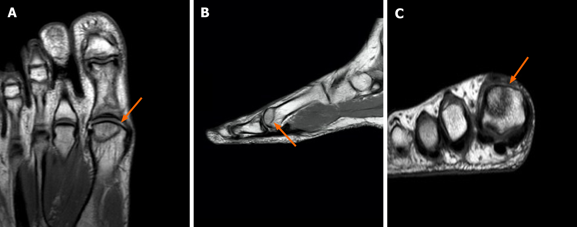
Figure 1 Initial foot magnetic resonance images.
A: Anterior; B: Sagittal; C: Coronal images. The images showing serpiginous line with bony oedema at distal phalangeal base and proximal phalangeal head on T1-weighted magnetic resonance images.
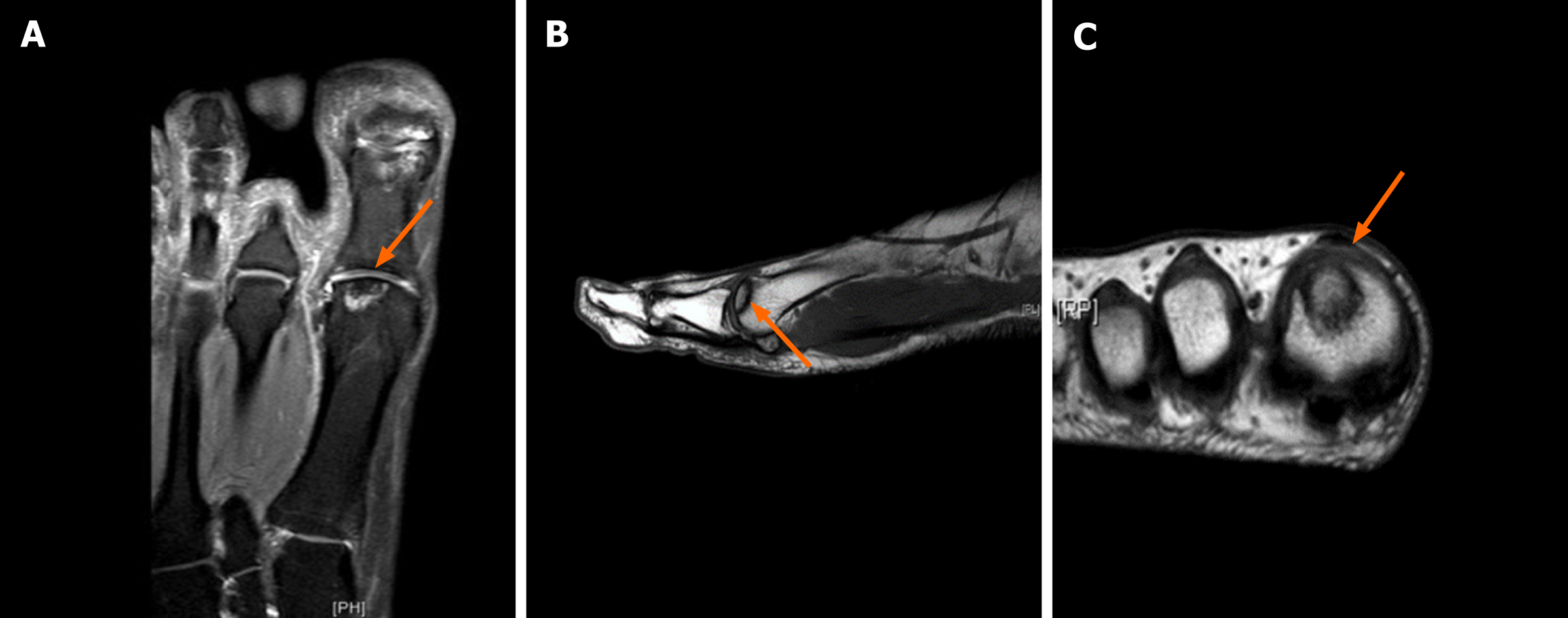
Figure 2 At follow-up after 14 mo.
A: Anterior; B: Sagittal; C: Coronal images. The images showing serpiginous line with reduced bony oedema at distal phalangeal base and proximal phalangeal head on T2-weighted magnetic resonance images.
- Citation: Siu RWH, Liu JHP, Man GCW, Ong MTY, Yung PSH. Avascular necrosis of the first metatarsal head in a young female adult: A case report and review of literature. World J Clin Cases 2021; 9(25): 7445-7452
- URL: https://www.wjgnet.com/2307-8960/full/v9/i25/7445.htm
- DOI: https://dx.doi.org/10.12998/wjcc.v9.i25.7445


