Copyright
©The Author(s) 2021.
World J Clin Cases. Sep 6, 2021; 9(25): 7381-7390
Published online Sep 6, 2021. doi: 10.12998/wjcc.v9.i25.7381
Published online Sep 6, 2021. doi: 10.12998/wjcc.v9.i25.7381
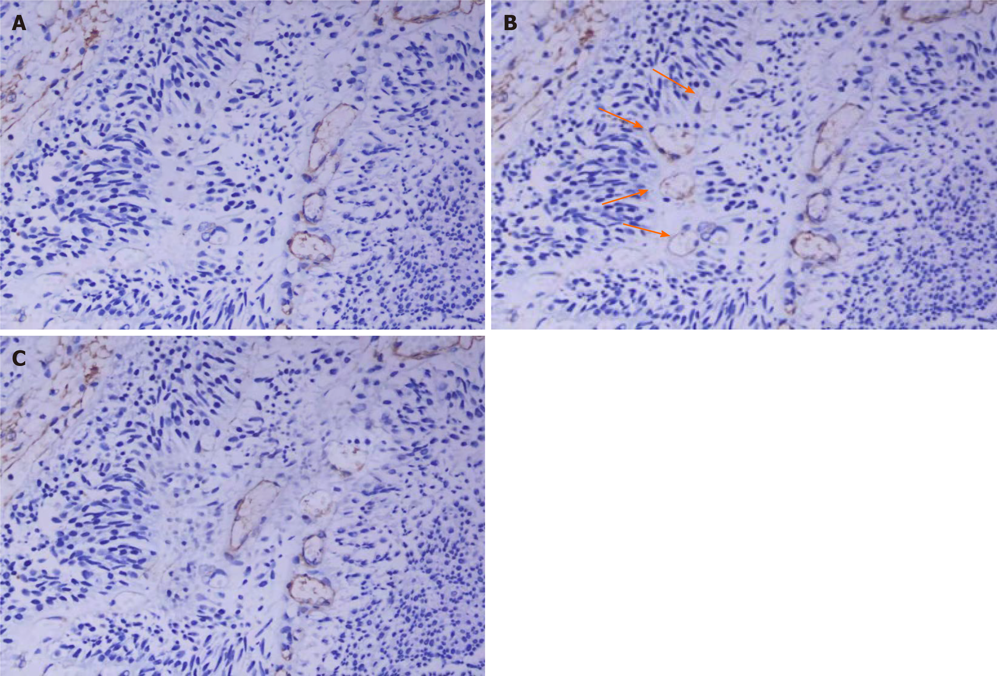
Figure 1 Two different types of microvessels were observed in bladder transitional cell carcinoma.
A: CD34-stained pictures clearly reveal differentiated blood vessels in bladder transitional cell carcinoma (× 400); B: Other blood vessels are stained with CD31 in the same position. As shown by the arrow position, undifferentiated blood vessels are not stained by the CD34 monoclonal antibody (× 400); C: Smooth muscle actin staining confirms that peripheral cells cover the blood vessels stained by CD34, but no peripheral cells cover the undifferentiated blood vessels in Figure 1B (× 400).
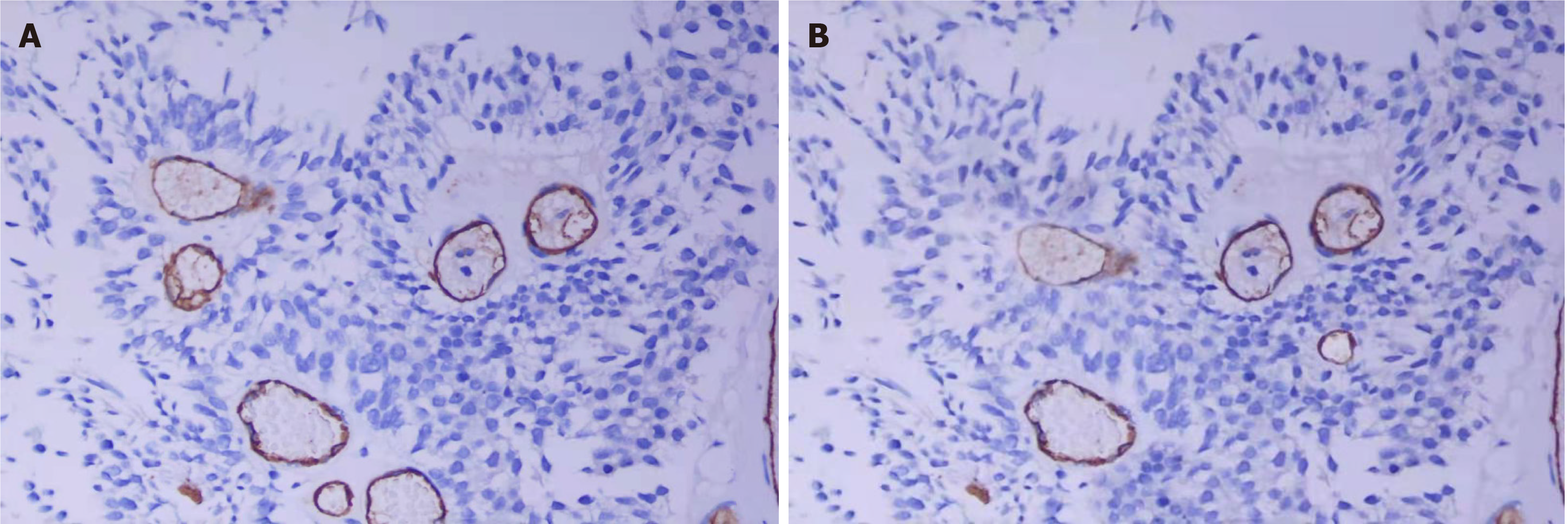
Figure 2 CD34 and CD105 stained pictures.
A: CD34 staining clearly revealed differentiated blood vessels in bladder transitional cell carcinoma (× 400); B: CD105 staining in the same position (× 400).
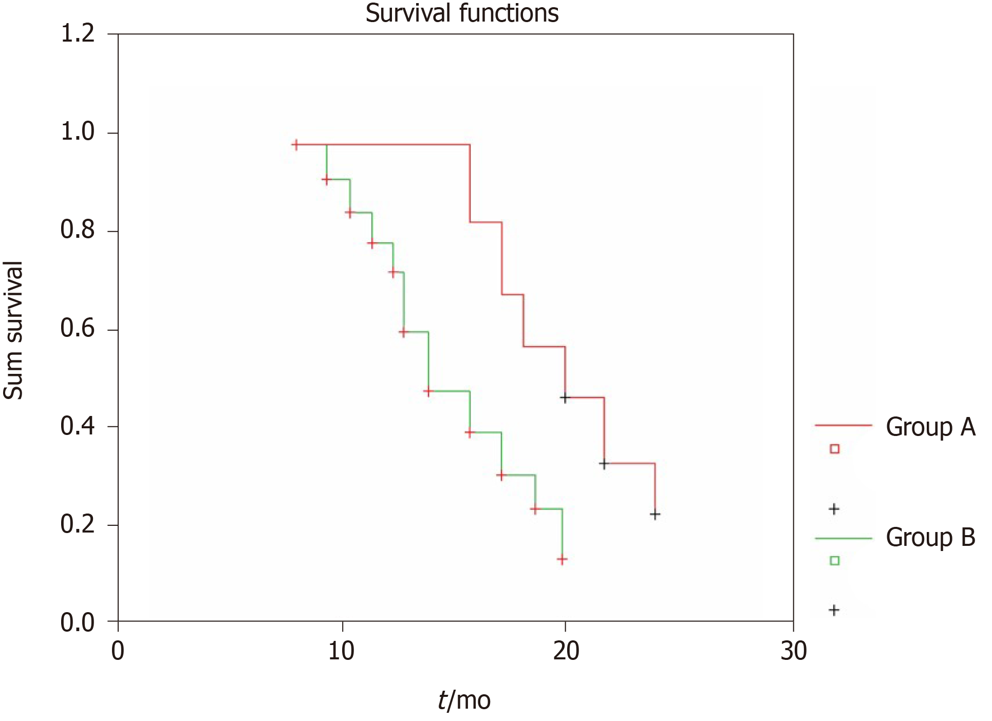
Figure 3 Kaplan-Meier tumor-free survival curve of bladder transitional cell carcinoma patients in the undifferentiated microvessel density groups.
- Citation: Wang HB, Qin Y, Yang JY. Research on the prognosis of different types of microvessels in bladder transitional cell carcinoma. World J Clin Cases 2021; 9(25): 7381-7390
- URL: https://www.wjgnet.com/2307-8960/full/v9/i25/7381.htm
- DOI: https://dx.doi.org/10.12998/wjcc.v9.i25.7381


