Copyright
©The Author(s) 2021.
World J Clin Cases. Sep 6, 2021; 9(25): 7330-7339
Published online Sep 6, 2021. doi: 10.12998/wjcc.v9.i25.7330
Published online Sep 6, 2021. doi: 10.12998/wjcc.v9.i25.7330
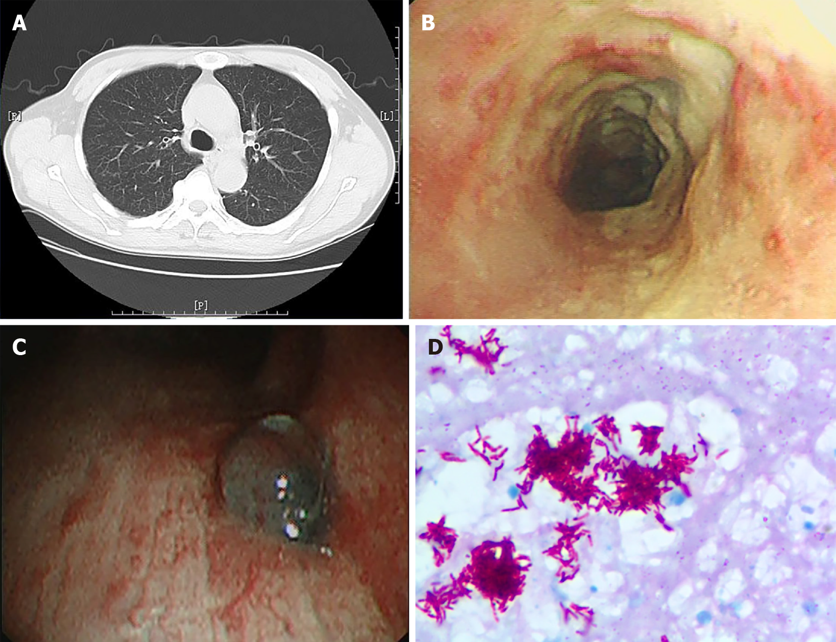
Figure 1 Chest computed tomography, bronchoscopy image and pathology image of a patient.
A: Chest computed tomography of a patient with tracheobronchial tuberculosis showing no obvious abnormalities; B: Bronchoscopy image showing white caseous necrotic tissue on tracheal wall; C: Bronchoscopy image showing granulomatous proliferation on the wall of lower right trachea; D: Necrotic granulomatosis was detected on the pathology image, accompanied with positive acid fast-staining and detection of mycobacterium tuberculosis complex, × 1000 magnification.
- Citation: Tang F, Lin LJ, Guo SL, Ye W, Zha XK, Cheng Y, Wu YF, Wang YM, Lyu XM, Fan XY, Lyu LP. Key determinants of misdiagnosis of tracheobronchial tuberculosis among senile patients in contemporary clinical practice: A retrospective analysis. World J Clin Cases 2021; 9(25): 7330-7339
- URL: https://www.wjgnet.com/2307-8960/full/v9/i25/7330.htm
- DOI: https://dx.doi.org/10.12998/wjcc.v9.i25.7330


