Copyright
©The Author(s) 2021.
World J Clin Cases. Aug 26, 2021; 9(24): 7269-7278
Published online Aug 26, 2021. doi: 10.12998/wjcc.v9.i24.7269
Published online Aug 26, 2021. doi: 10.12998/wjcc.v9.i24.7269
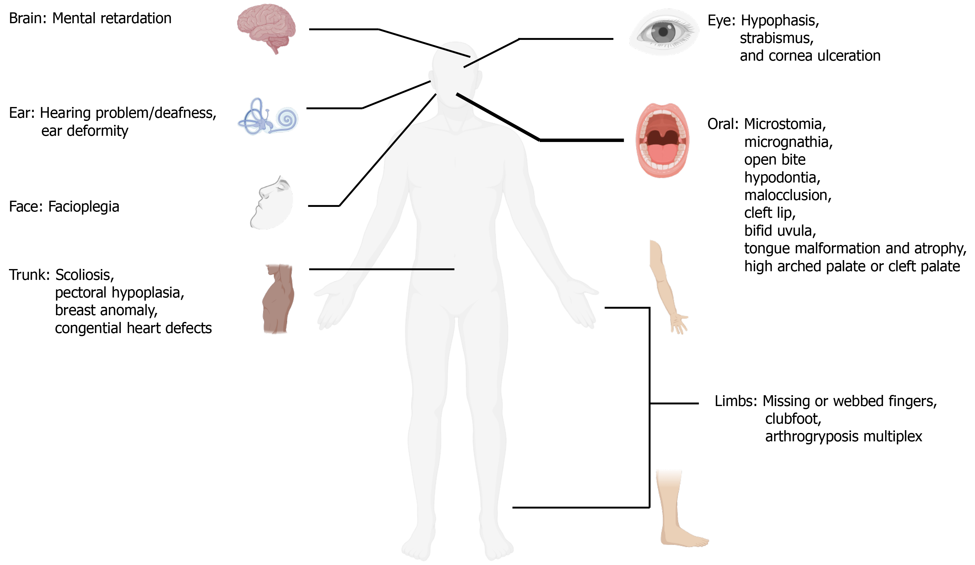
Figure 1 Clinical manifestations of Moebius syndrome.
Art adapted from BioRender.
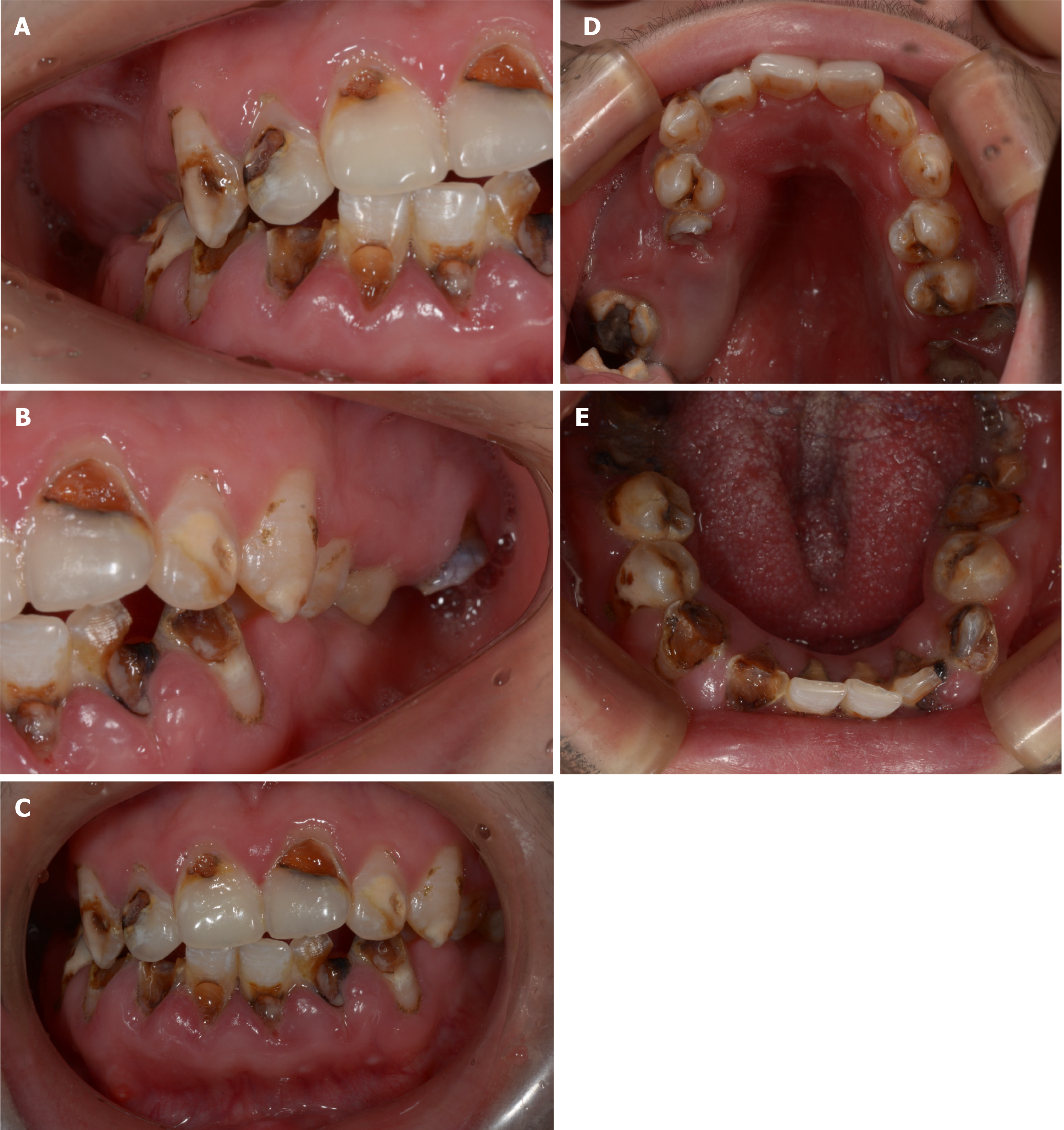
Figure 2 Pretreatment intraoral photographs.
A: Right lateral view; B: Left lateral view; C: Frontal view; D: Occlusal view of the maxillary arch; E: Occlusal view of the mandibular arch.
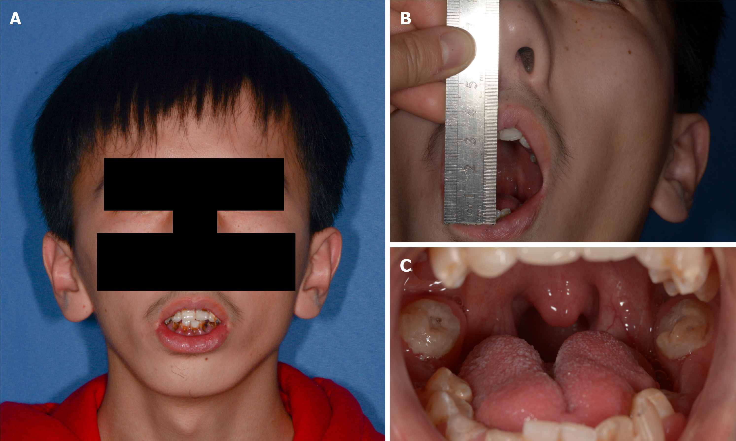
Figure 3 Extraoral facial photographs.
A: Frontal photograph; B: Limited mouth opening; C: Tongue malformation.
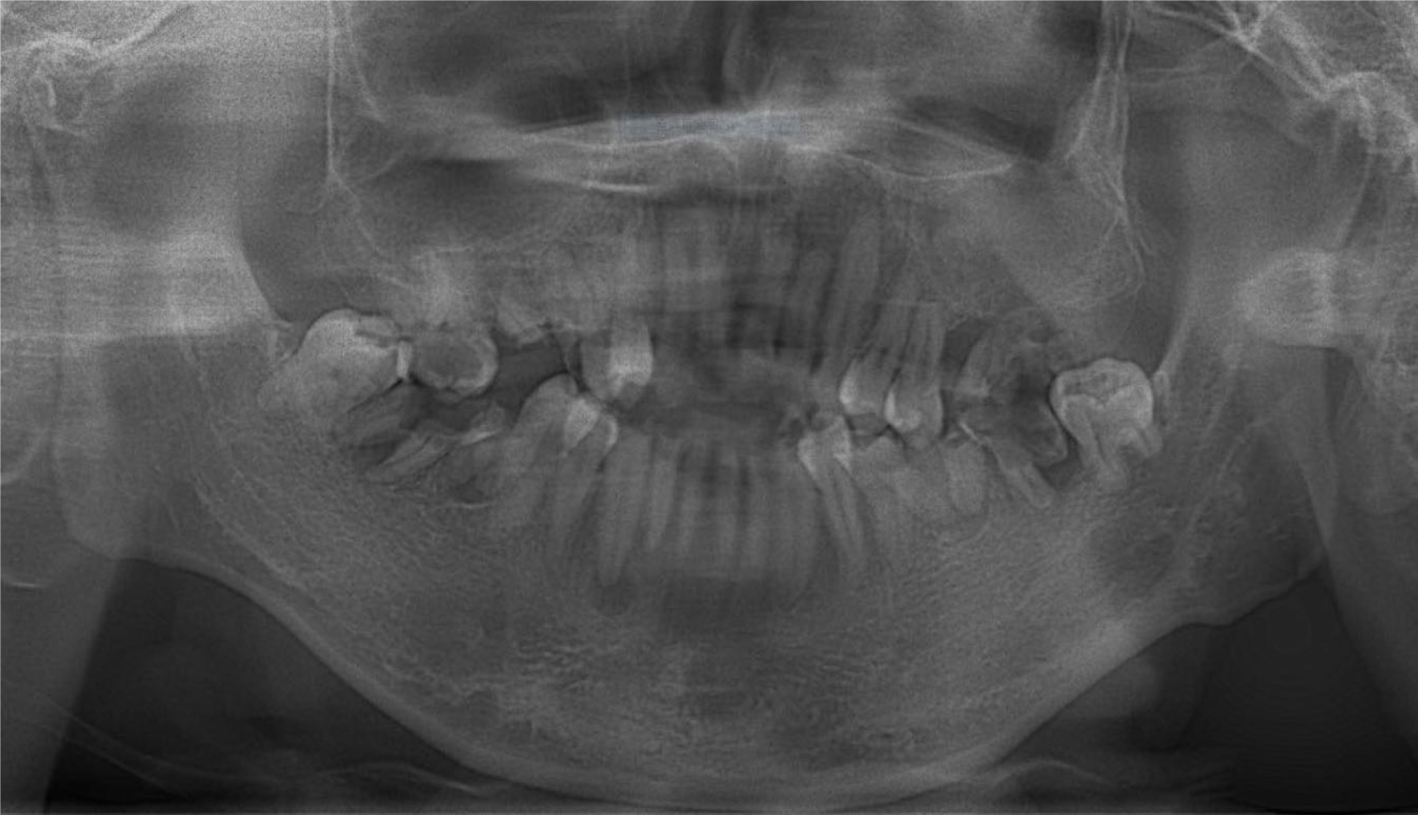
Figure 4 Initial panoramic image (2019).
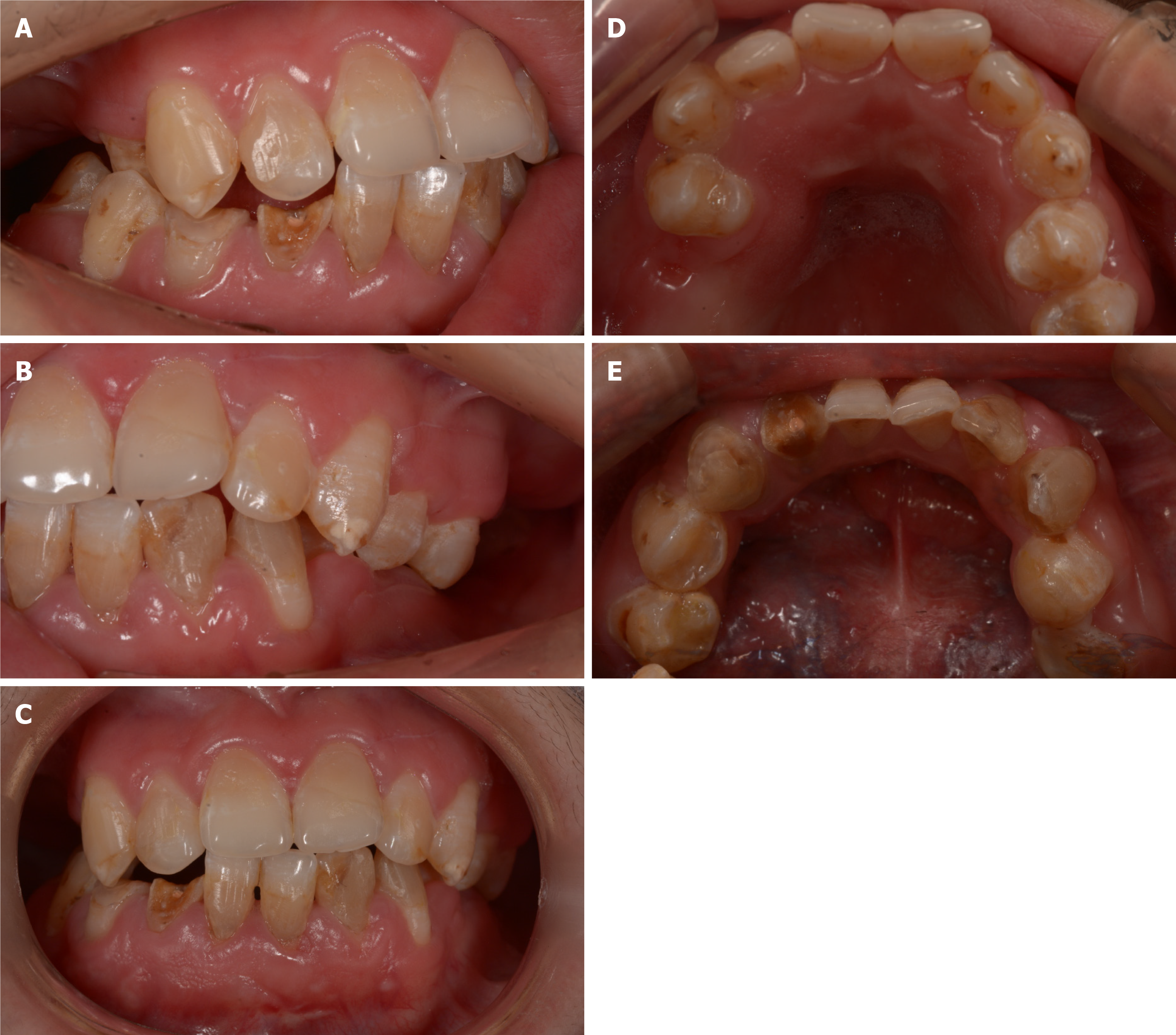
Figure 5 Posttreatment intraoral photographs.
A: Right lateral view; B: Left lateral view; C: Frontal view; D: Occlusal view of the maxillary arch; E: Occlusal view of the mandibular arch.
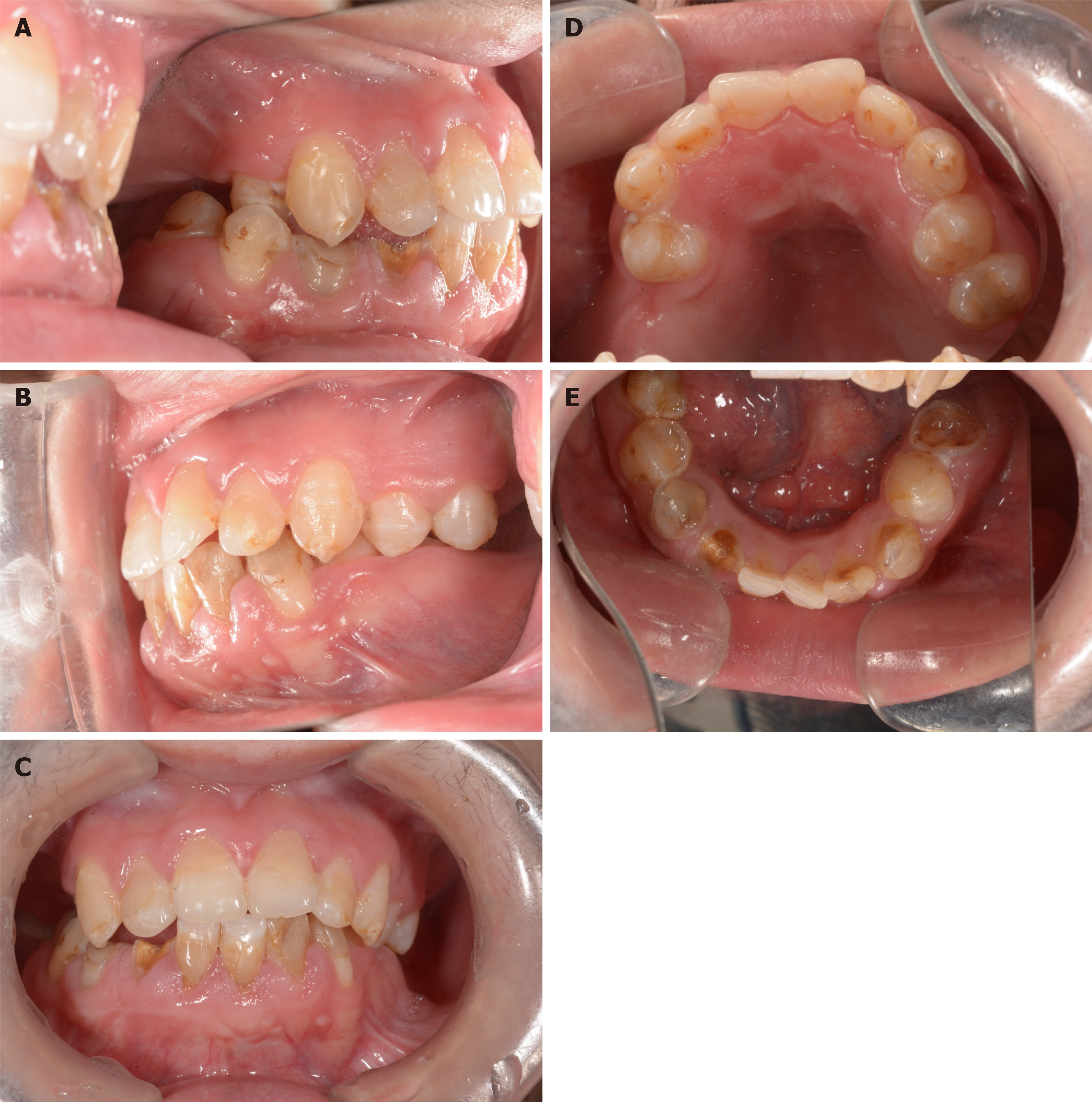
Figure 6 Two-year follow-up photographs.
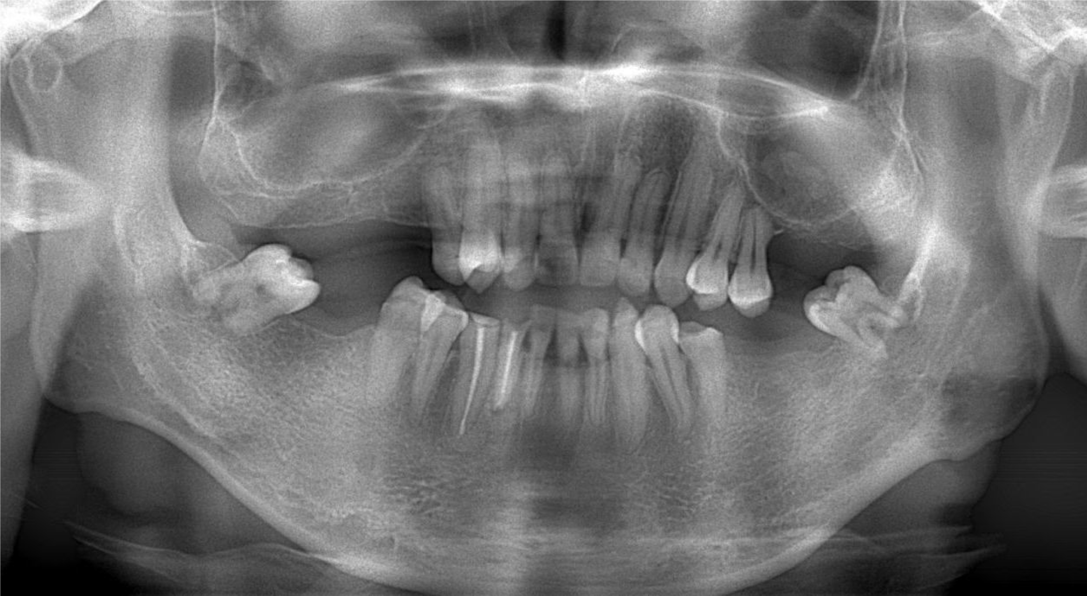
Figure 7 Two-year follow-up panoramic image (2021).
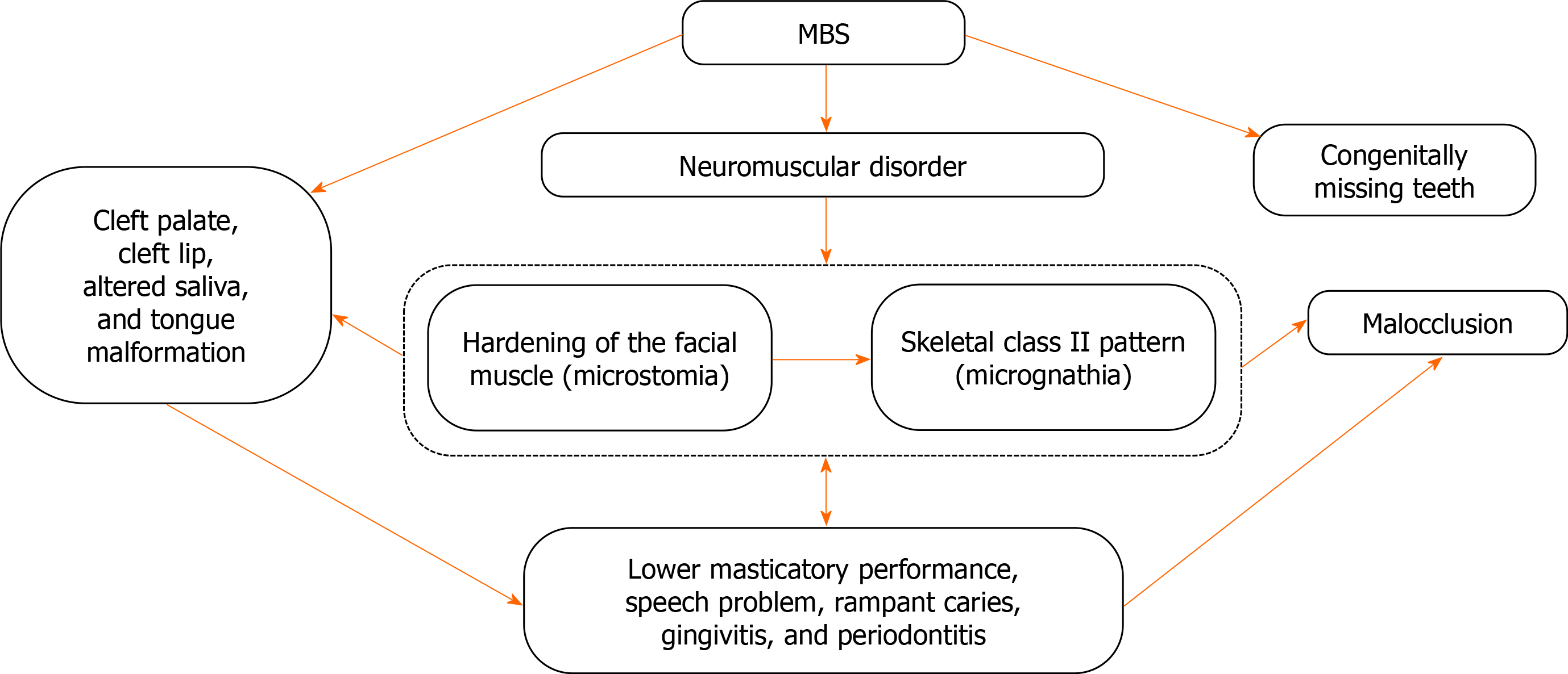
Figure 8 Interactions among oral complications of Moebius syndrome.
MBS: Moebius syndrome.
- Citation: Chen B, Li LX, Zhou LL. Dental management of a patient with Moebius syndrome: A case report. World J Clin Cases 2021; 9(24): 7269-7278
- URL: https://www.wjgnet.com/2307-8960/full/v9/i24/7269.htm
- DOI: https://dx.doi.org/10.12998/wjcc.v9.i24.7269


