Copyright
©The Author(s) 2021.
World J Clin Cases. Aug 26, 2021; 9(24): 7261-7268
Published online Aug 26, 2021. doi: 10.12998/wjcc.v9.i24.7261
Published online Aug 26, 2021. doi: 10.12998/wjcc.v9.i24.7261
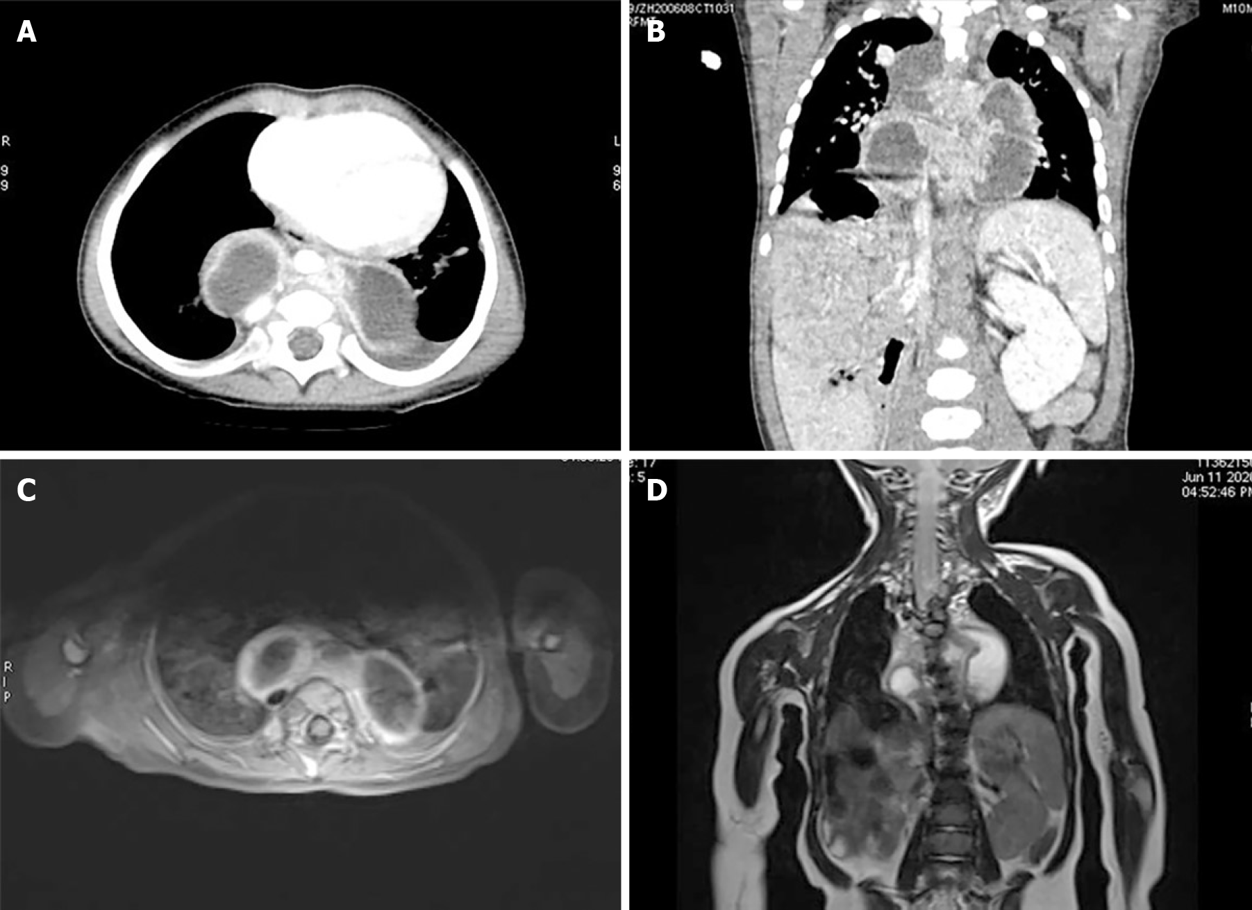
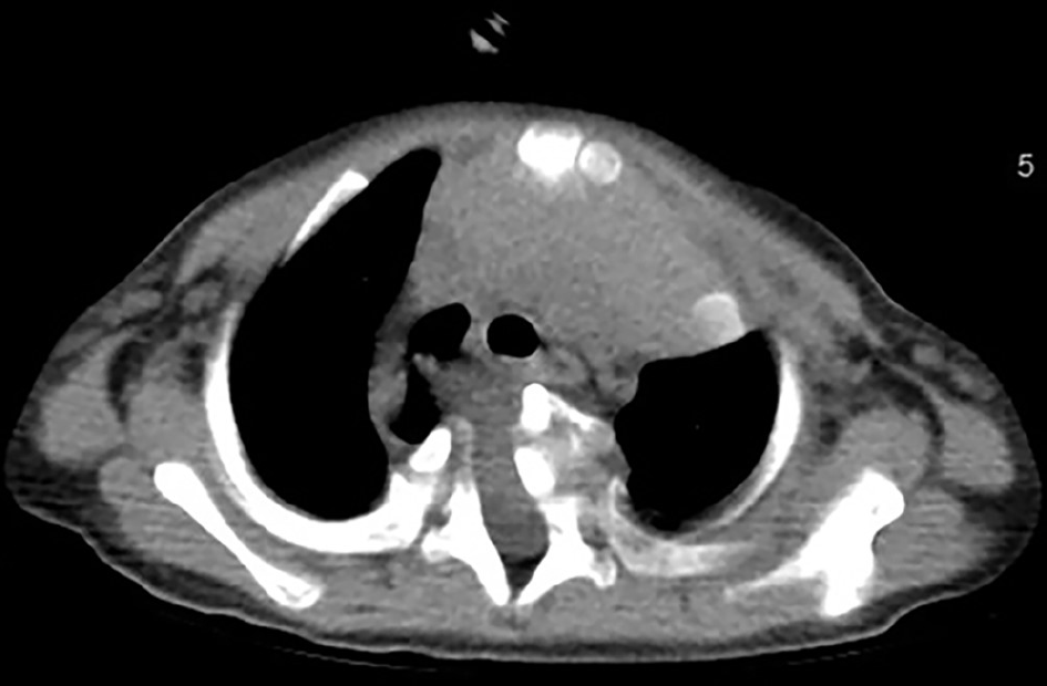
Figure 1 Imaging findings of posterior mediastinal cystic lesions.
A: Transected view of enhanced chest computed tomography showing posterior mediastinal cystic lesions; B: Sagittal view showing cystic lesions and myelomeningocele; C and D: Transected and sagittal view of enhanced chest magnetic resonance imaging.
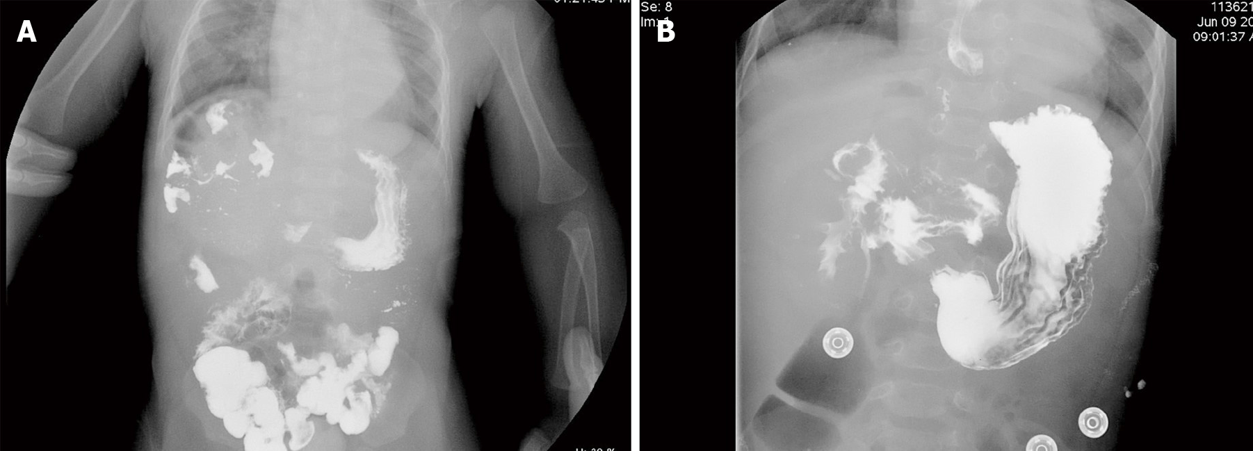
Figure 2 Upper gastroenterography showing a malrotated duodenum.
A: Locally the duodenal circle showed an abnormal shape and walked to the right upper abdomen; B: Anteroposterior radiograph of the abdomen.
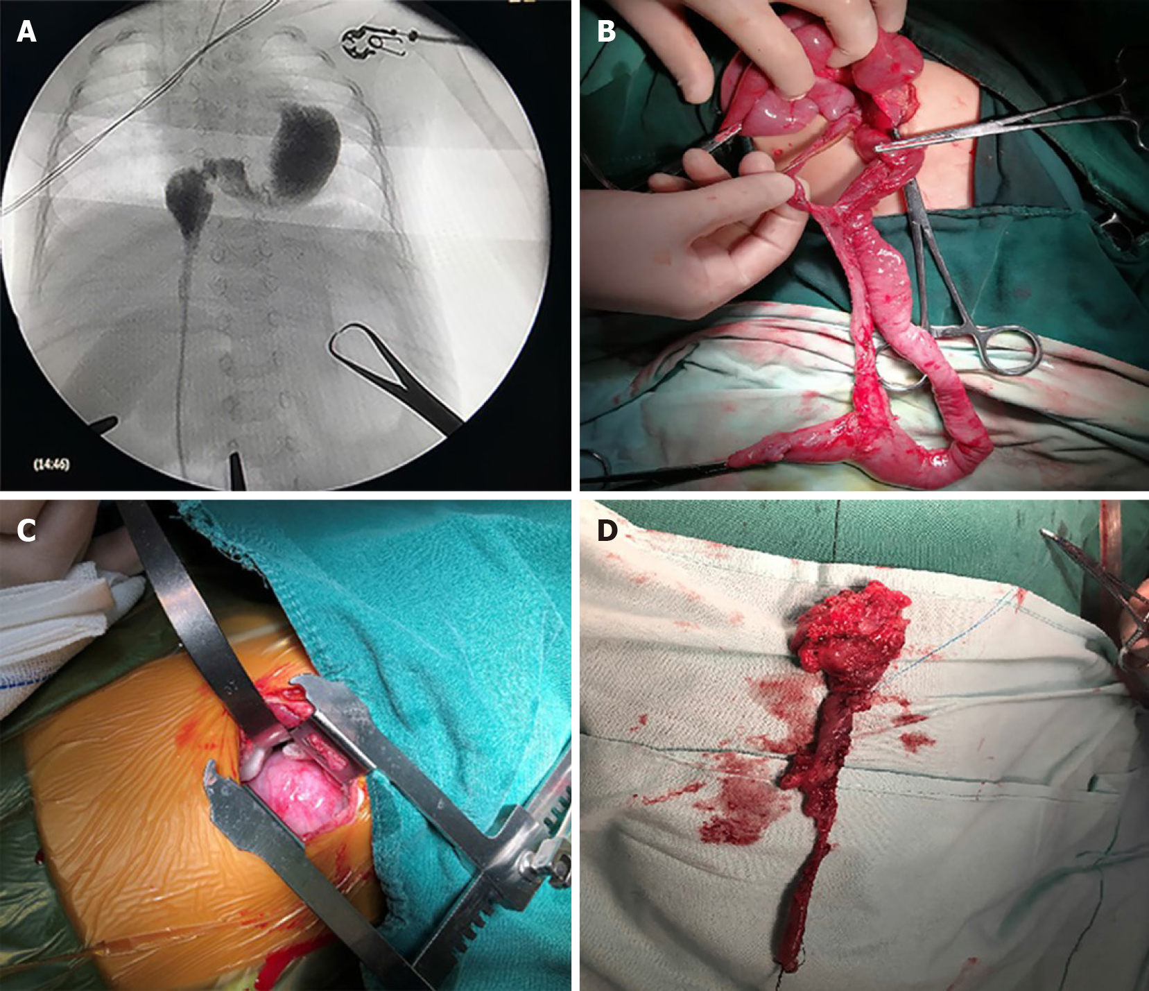
Figure 3 Intraoperative findings.
A: Intraoperative angiography showed that the intestinal tube passed through the diaphragm, extended upward to the posterior mediastinum, and crossed the midline right to left; B: The duplicated intestine and parallel intestinal tube were excised; C: An incision was made on the lateral posterior side of the left chest and intestinal tissue was found in the posterior mediastinum; D: Thoracic intestine was excised.
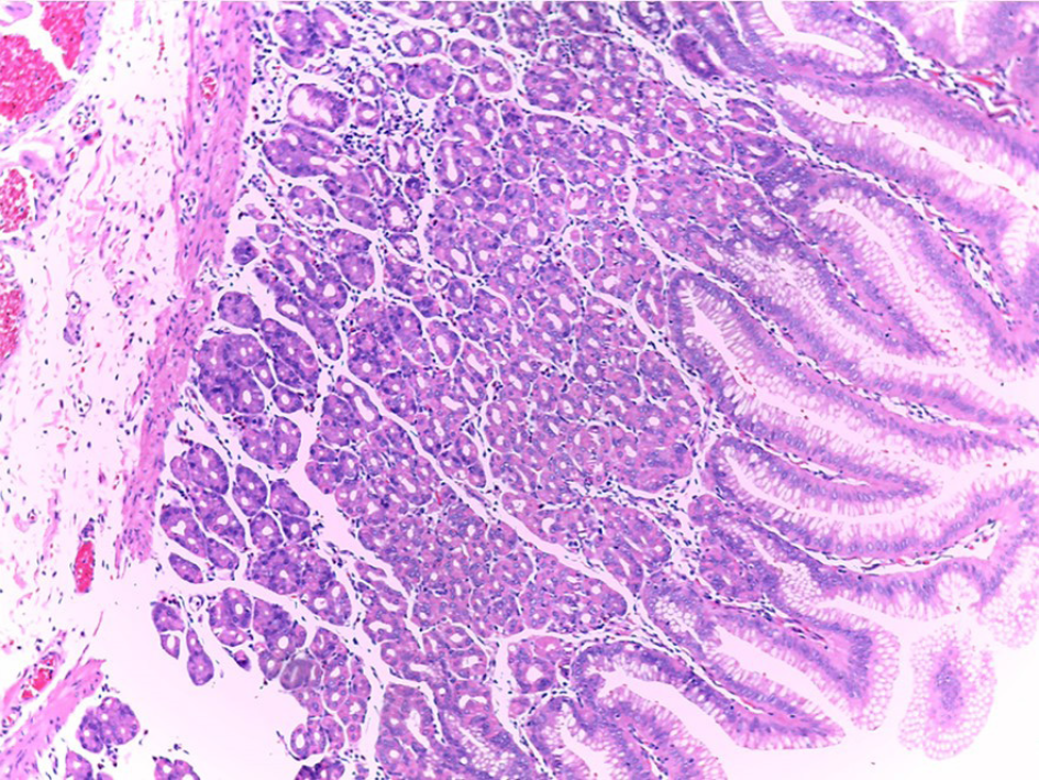
Figure 4 Pathological finding.
The mucosa is lined by intestinal epithelium and is composed of gastric-type glands (hematoxylin-eosin stain × 100).
Figure 5
Postoperative chest computed tomography showed myelomeningocele while the posterior mediastinal cyst was significantly reduced during outpatient follow-up.
- Citation: Yang SB, Yang H, Zheng S, Chen G. Thoracoabdominal duplication with hematochezia as an onset symptom in a baby: A case report. World J Clin Cases 2021; 9(24): 7261-7268
- URL: https://www.wjgnet.com/2307-8960/full/v9/i24/7261.htm
- DOI: https://dx.doi.org/10.12998/wjcc.v9.i24.7261


